. 2011 Apr 1;286(13):11456-68.
doi: 10.1074/jbc.M110.182881. Epub 2011 Feb 4.

CK2 phosphorylation of an acidic Ser/Thr di-isoleucine motif in the Na+/H+ exchanger NHE5 isoform promotes association with beta-arrestin2 and endocytosis

Affiliations

CK2 phosphorylation of an acidic Ser/Thr di-isoleucine motif in the Na+/H+ exchanger NHE5 isoform promotes association with beta-arrestin2 and endocytosis

Viktoria Lukashova et al. J Biol Chem. .

Abstract

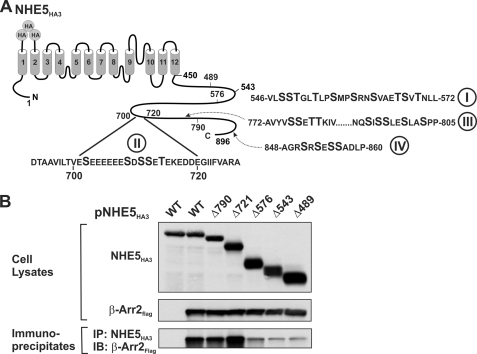
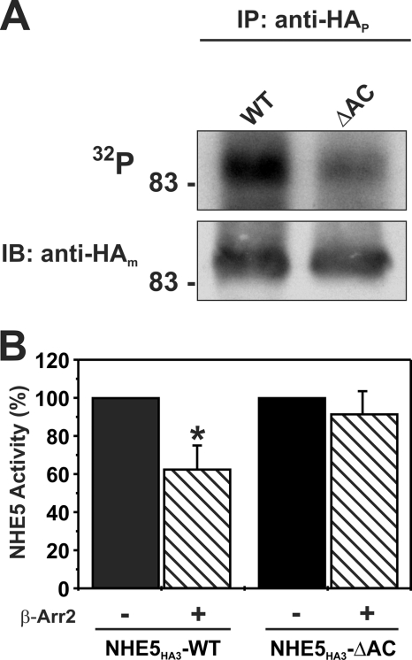
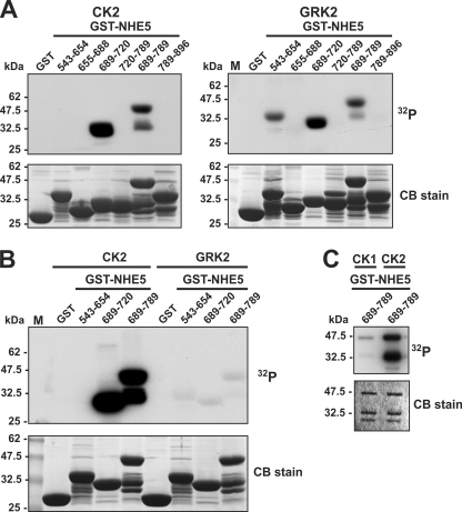
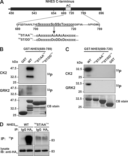
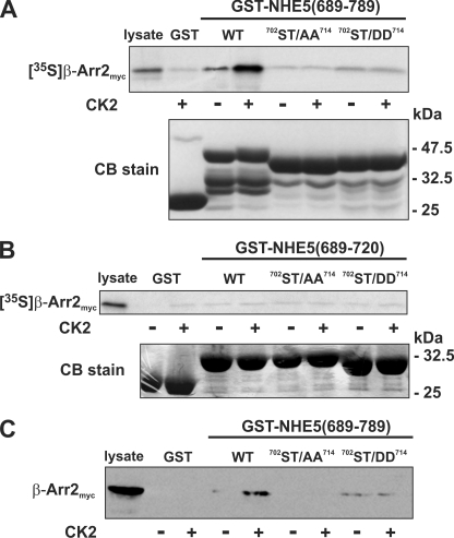
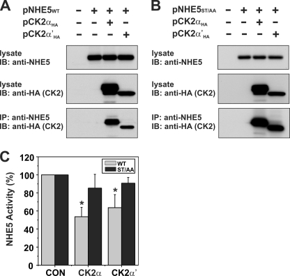
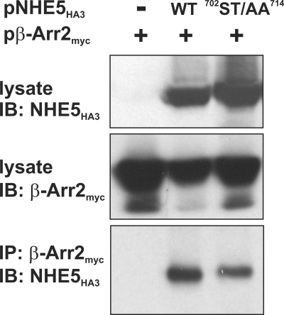
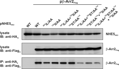
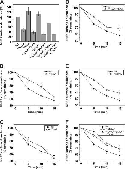
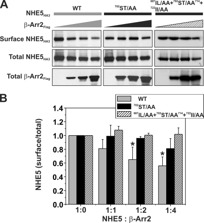
Internalization of the Na(+)/H(+) exchanger NHE5 into recycling endosomes is enhanced by the endocytic adaptor proteins β-arrestin1 and -2, best known for their preferential recognition of ligand-activated G protein-coupled receptors (GPCRs). However, the mechanism underlying their atypical association with non-GPCRs, such as NHE5, is unknown. In this study, we identified a highly acidic, serine/threonine-rich, di-isoleucine motif (amino acids 697-723) in the cytoplasmic C terminus of NHE5 that is recognized by β-arrestin2. Gross deletions of this site decreased the state of phosphorylation of NHE5 as well as its binding and responsiveness to β-arrestin2 in intact cells. More refined in vitro analyses showed that this site was robustly phosphorylated by the acidotropic protein kinase CK2, whereas other kinases, such as CK1 or the GPCR kinase GRK2, were considerably less potent. Simultaneous mutation of five Ser/Thr residues within 702-714 to Ala ((702)ST/AA(714)) abolished phosphorylation and binding of β-arrestin2. In transfected cells, the CK2 catalytic α subunit formed a complex with NHE5 and decreased wild-type but not (702)ST/AA(714) NHE5 activity, further supporting a regulatory role for this kinase. The rate of internalization of (702)ST/AA(714) was also diminished and relatively insensitive to overexpression of β-arrestin2. However, unlike in vitro, this mutant retained its ability to form a complex with β-arrestin2 despite its lack of responsiveness. Additional mutations of two di-isoleucine-based motifs (I697A/L698A and I722A/I723A) that immediately flank the acidic cluster, either separately or together, were required to disrupt their association. These data demonstrate that discrete elements of an elaborate sorting signal in NHE5 contribute to β-arrestin2 binding and trafficking along the recycling endosomal pathway.


Figures

FIGURE 1.
FIGURE 1.
Delineation of the β-arrestin2-binding domain in the C terminus of NHE5. A, schematic representation of the topological organization of NHE5 and locations of clusters of putative Ser/Thr phosphoacceptor sites in its cytoplasmic C terminus. B, full-length and C-terminal truncations of NHE5HA3 were transiently co-transfected with β-Arr2FLAG in CHO cells. NHE5HA3 was immunoprecipitated (IP) with a monoclonal anti-HA antibody, and the immunoprecipitates were resolved by SDS-PAGE followed by immunoblotting (IB) with a polyclonal anti-FLAG antibody to detect β-Arr2FLAG. Data shown are representative of at least three independent experiments.
FIGURE 2.
FIGURE 2.
An acidic Ser/Thr cluster in NHE5 confers responsiveness to β-Arr2 in intact cells. A, AP-1 cells stably expressing WT or mutant NHE5HA3 lacking the acidic cluster (amino acids 700–720; ΔAC) were metabolically labeled with [32P]orthophosphate, immunoprecipitated (IP) with a rabbit polyclonal anti-HA antibody (anti-HAp), and separated by SDS-PAGE. A representative autoradiograph is shown (top). Whole cell extracts were blotted (IB) with a mouse monoclonal anti-HA antibody (anti-HAm) (bottom). B, AP-1 cells were transiently co-transfected with NHE5HA3 WT or NHE5HA3-AC and β-Arr2myc (1:2 DNA ratio), and plasmalemmal NHE5 activity was measured 24 h post-transfection as described under “Experimental Procedures.” Values are the means ± S.D. (error bars) of at least three or four independent experiments (*, p < 0.05).
FIGURE 3.
FIGURE 3.
Comparison of in vitro phosphorylation of C-terminal segments of NHE5 by protein kinases CK1, CK2 and GRK2. A–C, purified GST and GST-NHE5 fusion proteins encoding peptide segments that span the cytoplasmic C terminus of NHE5 were incubated in vitro with 200 μm ATP and 1 μCi of [γ-32P]ATP and purified protein kinase CK1, CK2, or GRK2 as described under “Experimental Procedures.” The proteins in the reaction solution were resolved by SDS-PAGE. Radioactivity was detected using a PhosphorImager, and protein loading was visualized by staining with Coomassie Blue (CB) dye. Data shown are representative of at least three independent experiments.
FIGURE 4.
FIGURE 4.
Mutation of the acidic cluster abolishes NHE5 phosphorylation in vitro and in intact cells. A, schematic representation of the Ser and Thr residues in the acidic cluster (AC) that were mutated to either Ala (ST/AA) or Asp (ST/DD). B and C, purified GST-NHE5 fusion proteins containing amino acids 689–789 (B) or 689–720 (C) were phosphorylated in vitro by purified CK2 or GRK2 in the presence of 200 μm ATP and 1 μCi of [γ-32P]ATP, as described under “Experimental Procedures.” The reaction mixtures were fractionated by SDS-PAGE, and radioactivity and protein levels were detected using a PhosphorImager and Coomassie Blue (CB) dye, respectively. D, AP-1 cells stably expressing full-length NHE5HA3 WT or 702ST/AA714 mutant were metabolically labeled with [32P]orthophosphate, immunoprecipitated (IP) with control rabbit serum IgG or with a rabbit polyclonal anti-HA antibody (anti-HAp), and separated by SDS-PAGE. A representative autoradiograph is shown (top). Whole cell extracts were blotted (IB) with a mouse monoclonal anti-HA antibody (anti-HAm) (bottom). Data shown are representative of at least three independent experiments.
FIGURE 5.
FIGURE 5.
Role of phosphorylation in the in vitro binding of β-arrestin2 to the C terminus of NHE5. A–C, purified WT and mutant (ST/AA and ST/DD) constructs of GST-NHE5(689–789) (A and C) and GST-NHE5(689–720) (B) were preincubated in the absence or presence of purified CK2 and ATP (200 μm). The GST-NHE5 fusion proteins were then incubated in the presence of in vitro synthesized 35S-labeled β-Arr2myc (A and B) or CHO cell lysates containing β-Arr2myc (C). The GST complexes were purified using glutathione-agarose beads and resolved by SDS-PAGE. To measure binding of β-Arr2myc, the gels containing radiolabeled β-Arr2myc were dried, and the signals were detected using a PhosphorImager, whereas those containing non-radioactive β-Arr2myc were subject to immunoblotting. Parallel gels were stained with Coomassie Blue (CB) dye to assess protein loading. Data shown are representative of at least three independent experiments.
FIGURE 6.
FIGURE 6.
Catalytic α/α′ subunits of protein kinase CK2 form a complex with NHE5 and regulate its activity. Full-length NHE5 WT (A) and mutant (ST/AA) (B) constructs were transiently transfected alone or together with CK2αHA or CK2α′HA in Chinese hamster ovary AP-1 cells. Cells were lysed after 24 h of transfection. Aliquots of the lysates were removed for Western blot analysis of total cellular expression of NHE5 and CK2αHA or CK2α′HA, whereas the remainder was incubated with a rabbit polyclonal anti-NHE5 antibody to precipitate NHE5-containing protein complexes. The immunoprecipitates (IP) were fractionated by SDS-PAGE, followed by immunoblotting (IB) with a monoclonal anti-HA antibody to detect CK2αHA or CK2α′HA. C, AP-1 cells stably expressing NHE5WT or NHE5ST/AA were transiently transfected with CK2αHA or CK2α′HA. Twenty-four h post-transfection, plasmalemmal NHE5 activity was measured as the rate of amiloride-inhibitable, H+-activated 22Na+ influx, as described under “Experimental Procedures.” Values are the means ± S.D. (error bars) of four independent experiments (*, p < 0.05).
FIGURE 7.
FIGURE 7.
β-Arrestin2 forms a complex with wild-type and ST/AA mutant NHE5 in transfected cells. Chinese hamster ovary AP-1 cells were transiently transfected with expression plasmids containing β-Arr2myc alone or in combination with WT or mutant (ST/AA) NHE5HA3. After 24 h of transfection, cells were lysed, and β-Arr2myc was immunoprecipitated (IP) using a polyclonal anti-myc antibody. The immunoprecipitates were resolved by SDS-PAGE and analyzed by immunoblotting (IB) with a monoclonal anti-HA antibody.
FIGURE 8.
FIGURE 8.
Role of di-isoleucine motifs in β-Arr2 binding to NHE5. Chinese hamster ovary AP-1 cells were transiently cotransfected with expression plasmids containing β-Arr2FLAG or empty vector combined with WT or mutant variants of NHE5HA3. After transfection (24 h), cells were lysed, and aliquots were removed for Western blot analysis of total cellular expression of NHE5HA3 and β-Arr2FLAG. The remainder of the lysates were incubated with a rabbit polyclonal anti-HA antibody to precipitate NHE5-containing protein complexes. The immunoprecipitates (IP) were resolved by SDS-PAGE and analyzed by immunoblotting (IB) with a monoclonal anti-FLAG antibody. Data shown are representative of at least three separate experiments.
FIGURE 9.
FIGURE 9.
Measurement of cell surface abundance and rates of endocytosis of wild-type and mutant variants of NHE5. Cell surface abundance (A) and internalization of plasmalemmal wild-type and mutant variants of NHE5FLAG3 (B–F) as a function of time were measured using an ELISA as described under “Experimental Procedures.” The turnover of plasma membrane NHE5FLAG3 was monitored by the disappearance of cell surface bound anti-FLAG antibody in transiently transfected HEK-293 GripTiteTM MSR cells. Briefly, cells were labeled with a monoclonal anti-FLAG antibody on ice for 1 h and then chased over a 15-min period at 37 °C. The anti-FLAG antibody remaining at the cell surface was measured by fluorescence in the presence of a mouse HRP-conjugated secondary antibody and Amplex Red, as described under “Experimental Procedures.” The fluorescence was normalized for cellular proteins. The values are expressed as a percentage of the initial amount of NHE5 at the cell surface at time 0 min and represent the mean ± S.E. (error bars) of 4–6 independent experiments.
FIGURE 10.
FIGURE 10.
Phosphorylation of the C-terminal acidic Ser/Thr cluster is important for β-arrestin2-mediated internalization of NHE5. A, AP-1 cells in 10-cm dishes were transiently cotransfected (24 h) with 10 μg of cDNA containing a fixed amount (2 μg) of WT or mutant (702ST/AA714 or 697IL/AA + 702ST/AA714 + 722II/AA) NHE5HA3 and increasing amounts (0–8 μg) of β-Arr2FLAG. The total DNA/transfection was held constant at 10 μg/dish by adjusting with empty parental vector pCMV. Cell surface NHE5 protein was isolated using a cell surface biotinylation assay (see “Experimental Procedures”) and compared with total cellular levels of NHE5 (10% of lysate). The fractions were resolved by SDS-PAGE and immunoblotted using antibodies to the respective epitope tags, and the intensities of the bands were quantified by densitometry. B, for comparative purposes, the ratio of surface/total NHE5 for cells expressing NHE5HA alone (control) was normalized to a value of 1. Values are the means ± S.E. (error bars) of at least four independent experiments (*, p < 0.05).

Similar articles

Cited by

References

    1. Brett C. L., Donowitz M., Rao R. (2005) Am. J. Physiol. Cell Physiol. 288, C223–C239 - PubMed
    1. Casey J. R., Grinstein S., Orlowski J. (2010) Nat. Rev. Mol. Cell Biol. 11, 50–61 - PubMed
    1. Wang D., King S. M., Quill T. A., Doolittle L. K., Garbers D. L. (2003) Nat. Cell Biol. 5, 1117–1122 - PubMed
    1. Wang D., Hu J., Bobulescu I. A., Quill T. A., McLeroy P., Moe O. W., Garbers D. L. (2007) Proc. Natl. Acad. Sci. U.S.A. 104, 9325–9330 - PMC - PubMed
    1. Bobulescu I. A., Moe O. W. (2006) Semin. Nephrol. 26, 334–344 - PMC - PubMed

Publication types

MeSH terms