Review
. 2011 Apr;10(4):466-73.
doi: 10.1128/EC.00313-10. Epub 2011 Feb 4.

Cell signals, cell contacts, and the organization of yeast communities

Affiliations
Review

Cell signals, cell contacts, and the organization of yeast communities

Saul M Honigberg. Eukaryot Cell. 2011 Apr.

Abstract

Even relatively simple species have evolved mechanisms to organize individual organisms into communities, such that the fitness of the group is greater than the fitness of isolated individuals. Within the fungal kingdom, the ability of many yeast species to organize into communities is crucial for their growth and survival, and this property has important impacts both on the economy and on human health. Over the last few years, studies of Saccharomyces cerevisiae have revealed several fundamental properties of yeast communities. First, strain-to-strain variation in the structures of these groups is attributable in part to variability in the expression and functions of adhesin proteins. Second, the extracellular matrix surrounding these communities can protect them from environmental stress and may also be important in cell signaling. Finally, diffusible signals between cells contribute to community organization so that different regions of a community express different genes and adopt different cell fates. These findings provide an arena in which to view fundamental mechanisms by which contacts and signals between individual organisms allow them to assemble into functional communities.


Figures

Fig. 1.
Fig. 1.
Patterns of differentiation and gene expression in colonies. (A) Section from central region of a 6-day-old wild yeast colony. The top of the image is closest to the top of the colony. The region of the section indicated by the gray bar has a high frequency of sporulation, whereas the underlying region contains no asci. A representative ascus is indicated by an arrow. Scale bar, 50 μm. (Reprinted from reference with permission of the publisher.) (B) Pseudohyphal and meiotic differentiation on the surfaces of minicolonies. Scale bars, 100 μm (left panel) and 10 μm (right panel). (Reprinted from reference with permission of the publisher.) (C) Expression of ATO1-GFP within a colony. The upper panel is a side view obtained after slicing a 10-day-old colony in half. Scale bar, 150 μm. The lower panel is viewed from the bottom of a 10-day-old colony. Scale bar, 500 μm. (Reprinted from reference with permission of the publisher.) (D) Colony expression pattern of two meiotic genes. The upper panel is a colony containing the IME1 promoter fused to lacZ, and the lower panel is a colony containing the IME2 promoter fused to lacZ. (Reprinted from reference with permission of the publisher.)
Fig. 2.
Fig. 2.
Dependence of colony structure on strain background and growth conditions. (A) YJM311 (a clinical isolate of S. cerevisiae) (reprinted from reference with permission of the publisher); (B) PMY348 (reprinted from reference with permission of the publisher); (C) SH561 (SK1 background) grown on yeast extract-peptone-dextrose (YPD); (D) SH561 grown on YP-glycerol. Scale bars, 1 mm.
Fig. 3.
Fig. 3.
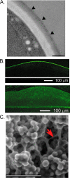
Roles of extracellular matrix (ECM) in community organization. (A) Flocs expressing FLO1 are coated with extracellular matrix, as viewed by transmission electron microscopy. Arrowheads indicate a gray staining region on the surface of a floc. (Reprinted from reference with permission of the publisher.) (B) Tight attachments at the colony surface. (Top) ConA-Alexa Fluor staining of the top of a colony; (bottom) ConA-Alexa Fluor staining of a sliced colony from the side. (Reprinted from reference with permission of the publisher.) (C) Scanning electron micrograph of a fluffy colony. A representative bridge of ECM is indicated by the arrow. Scale bar, 20 μm. (Reprinted from reference with permission of the publisher.)
Fig. 4.
Fig. 4.
Summary of interactions affecting yeast community organization. (A) Cell-ECM interactions. Mucins, such as Msb2p, and flocculins, such as Flo11p, that are secreted from cells help to establish the S. cerevisiae extracellular matrix (ECM) and may also signal other cells through this matrix. (B) Diffusible signals between cells. Alkaline pH sensed through the Rim101 pathway, ammonia/ammonium produced by the Ato transporters, and perhaps also reactive oxygen species are all diffusible signals which contribute to the organization of yeast into communities. (C) Cell-cell contacts. Flocculins are cell surface proteins required for contacts between cells. (D) Cell-surface contacts. Flocculins also required for contacts between cells and between cells and surfaces.

Similar articles

Cited by

References

    1. Barrales R. R., Jimenez J., Ibeas J. I. 2008. Identification of novel activation mechanisms for FLO11 regulation in Saccharomyces cerevisiae. Genetics 178:145–156 - PMC - PubMed
    1. Bastidas R. J., Heitman J. 2009. Trimorphic stepping stones pave the way to fungal virulence. Proc. Natl. Acad. Sci. U. S. A. 106:351–352 - PMC - PubMed
    1. Beauvais A., Loussert C., Prevost M. C., Verstrepen K., Latge J. P. 2009. Characterization of a biofilm-like extracellular matrix in FLO1-expressing Saccharomyces cerevisiae cells. FEMS Yeast Res. 9:411–419 - PubMed
    1. Bedard K., Krause K. H. 2007. The NOX family of ROS-generating NADPH oxidases: physiology and pathophysiology. Physiol. Rev. 87:245–313 - PubMed
    1. Blankenship J. R., Mitchell A. P. 2006. How to build a biofilm: a fungal perspective. Curr. Opin. Microbiol. 9:588–594 - PubMed

Publication types

MeSH terms

LinkOut - more resources