. 2011;24(3):547-57.
doi: 10.3233/JAD-2011-101782.

Synaptic loss in the inferior temporal gyrus in mild cognitive impairment and Alzheimer's disease

Affiliations

Synaptic loss in the inferior temporal gyrus in mild cognitive impairment and Alzheimer's disease

Stephen W Scheff et al. J Alzheimers Dis. 2011.

Abstract

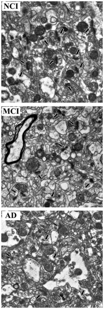
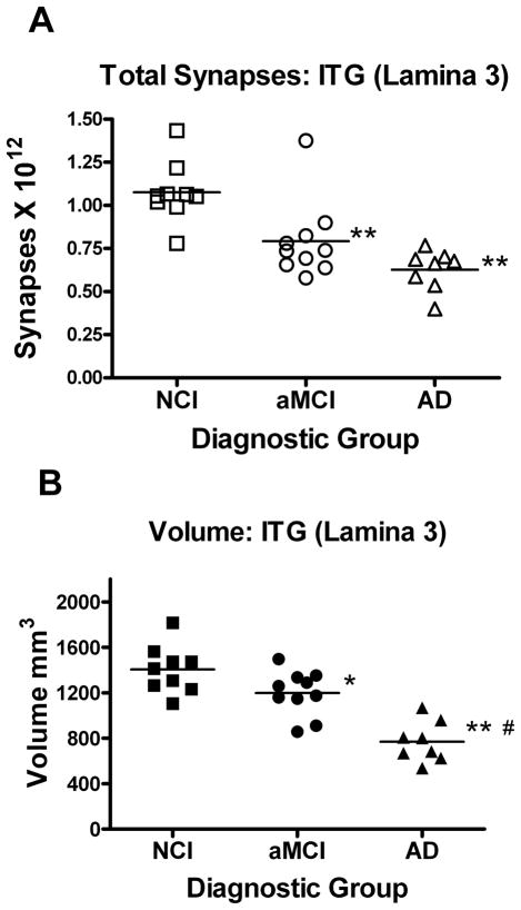
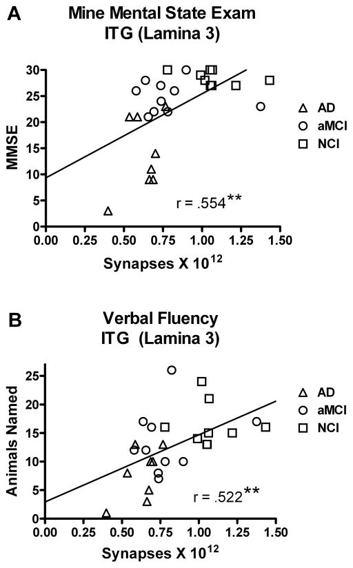
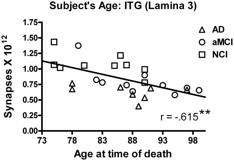
Alzheimer's disease (AD) is a slowly progressing form of dementia characterized in its earliest stages as a loss of memory. Individuals with amnestic mild cognitive impairment (aMCI) may be in the earliest stages of the disease and represent an opportunity to identify pathological changes related to the progression of AD. Synaptic loss is one of the hallmarks of AD and associated with cognitive impairment. The inferior temporal gyrus plays an important role in verbal fluency, a cognitive function affected early in the onset of AD. Unbiased stereology coupled with electron microscopy was used to quantify total synaptic numbers in lamina 3 of the inferior temporal gyrus from short postmortem autopsy tissue harvested from subjects who died at different cognitive stages during the progression of AD. Individuals with aMCI had significantly fewer synapses (36%) compared to individuals with no cognitive impairment. Individuals with AD showed a loss of synapses very similar to the aMCI cohort. Synaptic numbers correlated highly with Mini Mental State Examination scores and a test of category verbal fluency. These results demonstrate that the inferior temporal gyrus is affected during the prodromal stage of the disease and may underlie some of the early AD-related clinical dysfunctions.


Figures

Figure 1
Figure 1
Representative electron micrographs of lamina 3 of the inferior temporal gyrus showing synaptic complexes (arrows) in tissue from the three different clinical groups A) no cognitive impairment (NCI), B) amnestic mild cognitive impairment (MCI), C) Alzheimer disease (AD). In all tissue, the synaptic complexes appeared normal with synaptic vesicles observed in the presynaptic component and a synaptic density observed in the postsynaptic component. Calibration bar = 1.0 μm.
Figure 2
Figure 2
Estimate of the total number of synapses (A) in lamina 3 of the inferior temporal gyrus. Subjects were categorized clinically as no cognitive impairment (NCI), amnestic mild cognitive impairment (aMCI), or mild to moderate Alzheimer’s disease (AD). Estimates were obtained using unbiased stereology coupled with electron microscopic imaging of synapses. The total volume (B) of lamina 3 of the inferior temporal gyrus was estimated with the Cavalieri method directly from tissue sections immediately adjacent to regions used for synaptic counts. Single points represent individual subjects. Horizontal lines indicates group median. *p < 0.05; **p < 0.005 compared to NCI; #p < 0.005 compared to aMCI.
Figure 3
Figure 3
Relationship between estimates of total number of synapses in lamina 3 of the inferior temporal gyrus and the subjects score on the Mini Mental Status Examination (MMSE) (A) and a test of verbal fluency using an animal naming test (B). Subjects were categorized clinically as no cognitive impairment (NCI), amnestic mild cognitive impairment (aMCI), or mild to moderate Alzheimer’s disease (AD). As the total number of synapses increased, so did the subjects’ performance on these two cognitive tests. Lines are used to represent the direction of the association and do not indicate a line of regression. Single points represent individual subjects’ scores for each group. **p < 0.005
Figure 4
Figure 4
Relationship between estimates of the total number of synapses in lamina 3 of the inferior temporal gyrus and an individual’s age at the time of death. Subjects were categorized clinically as no cognitive impairment (NCI), amnestic mild cognitive impairment (aMCI), or mild to moderate Alzheimer’s disease (AD). There was a significant association with decreased synaptic numbers with increased subject’s age. Lines are used to represent the direction of the association and do not indicate a line of regression. Single points represent individual subjects’ scores for each group. **p < 0.005

References

    1. Weiner MF, Neubecker KE, Bret ME, Hynan LS. Language in Alzheimer's disease. J Clin Psychiatry. 2008;69:1223–7. - PMC - PubMed
    1. Barberger-Gateau P, Fabrigoule C, Helmer C, Rouch I, Dartigues JF. Functional impairment in instrumental activities of daily living: an early clinical sign of dementia? J Am Geriatr Soc. 1999;47:456–62. - PubMed
    1. Cahn DA, Salmon DP, Bondi MW, Butters N, Johnson SA, Wiederholt WC, Barrett-Connor E. A population-based analysis of qualitative features of the neuropsychological test performance of individuals with dementia of the Alzheimer type: implications for individuals with questionable dementia. J Int Neuropsychol Soc. 1997;3:387–93. - PubMed
    1. Chertkow H, Bub D. Semantic memory loss in dementia of Alzheimer's type. What do various measures measure? Brain. 1990;113 ( Pt 2):397–417. - PubMed
    1. Monsch AU, Bondi MW, Butters N, Salmon DP, Katzman R, Thal LJ. Comparisons of verbal fluency tasks in the detection of dementia of the Alzheimer type. Arch Neurol. 1992;49:1253–8. - PubMed

Publication types

Substances