. 2011 Mar 16;30(6):1054-66.
doi: 10.1038/emboj.2011.23. Epub 2011 Feb 4.

RNA content in the nucleolus alters p53 acetylation via MYBBP1A

Affiliations

RNA content in the nucleolus alters p53 acetylation via MYBBP1A

Takao Kuroda et al. EMBO J. .

Abstract

A number of external and internal insults disrupt nucleolar structure, and the resulting nucleolar stress stabilizes and activates p53. We show here that nucleolar disruption induces acetylation and accumulation of p53 without phosphorylation. We identified three nucleolar proteins, MYBBP1A, RPL5, and RPL11, involved in p53 acetylation and accumulation. MYBBP1A was tethered to the nucleolus through nucleolar RNA. When rRNA transcription was suppressed by nucleolar stress, MYBBP1A translocated to the nucleoplasm and facilitated p53-p300 interaction to enhance p53 acetylation. We also found that RPL5 and RPL11 were required for rRNA export from the nucleolus. Depletion of RPL5 or RPL11 blocked rRNA export and counteracted reduction of nucleolar RNA levels caused by inhibition of rRNA transcription. As a result, RPL5 or RPL11 depletion inhibited MYBBP1A translocation and p53 activation. Our observations indicated that a dynamic equilibrium between RNA generation and export regulated nucleolar RNA content. Perturbation of this balance by nucleolar stress altered the nucleolar RNA content and modulated p53 activity.


Conflict of interest statement

The authors declare that they have no conflict of interest.

Figures

Figure 1
Figure 1
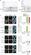
MYBBP1A is necessary for p53 acetylation induced by nucleolar disruption. (A) Knockdown of TIF-IA induces acetylation at multiple lysine residues in p53. MCF-7 cells were treated with siCont or siTIF-IA for 48 h, and the cell lysates were analysed by immunoblot using the indicated antibodies. (B) Nucleolar disruption releases the nucleolar protein MYBBP1A into the nucleoplasm. MCF-7 cells were treated with siCont or siTIF-IA for 48 h, and the localization of MYBBP1A and UBF was visualized by immunofluorescence using anti-MYBBP1A and anti-UBF antibodies. (C) Knockdown of MYBBP1A antagonizes the acetylation of p53 proteins caused by nucleolar disruption. MCF-7 cells were treated with the siCont or MYBBP1A siRNA (siMYBBP) with or without siTIF-IA for 48 h, and cell lysates were analysed by immunoblot using the indicated antibodies. (D) Knockdown of MYBBP1A decreases the acetylation levels at multiple lysine residues in the p53 protein. (Left) MCF-7 cells were treated with the siCont or two independent siMYBBPs (siMYBBP and siMYBBP-2) with or without siTIF-IA for 48 h, and cell lysates were analysed by immunoblot using site-specific acetylated p53 antibodies. (Right) Relative quantification of acetylation levels of the p53 protein. The intensities of the acetylated p53 proteins were corrected using the p53 protein level. The intensity of the siCont-treated cells was normalized to 1.0. (E) Knockdown of MYBBP1A antagonizes the acetylation of p53 induced by low-dose ActD treatment. MCF-7 cells were treated with siCont or siMYBBP for 48 h, followed by 5 nM ActD treatment for indicated times. The cell lysates were analysed by immunoblot using the indicated antibodies. (F) Recovery of the acetylation levels of p53 protein by introducing siRNA-resistant MYBBP1A along with siRNA for MYBBP1A. p53-deficient H1299 cells were treated with the siCont or siMYBBP with or without siTIF-IA for 24 h before transfection with Myc-p53 and siRNA-resistant MYBBP1A (MYBBP1A-simut). Twenty-four hours after transfection, cell lysates were analysed by immunoblot with the indicated antibodies.
Figure 2
Figure 2
MYBBP1A directly binds to p53, increasing the binding of p300 to p53. (A) Endogenous MYBBP1A associates with p53. MCF-7 cells were treated with siTIF-IA for 48 h. The cell lysates were immunoprecipitated with normal rabbit IgG or anti-MYBBP1A antibodies and analysed by immunoblot using antibodies against MYBBP1A and p53. (B) (Left) Domain structure of the full-length p53 and various deletion mutants. TAD, transactivation domain; PRD, proline-rich domain; DBD, DNA-binding domain; NLS, nuclear localization signal; TET, tetramerization domain; CRD, C-terminal regulatory domain; K, lysine residues that can be acetylated in the CRD region. (Right) MYBBP1A directly binds to the CRD region in p53. 35S-labelled MYBBP1A was incubated with the GST-fused full-length or truncated p53 proteins. After extensive washing, bound proteins were analysed by SDS–PAGE and autoradiography. (C) MYBBP1A forms a ternary complex with p53 and p300. MCF-7 cells were treated with siTIF-IA for 48 h. The cell lysates were sequentially immunoprecipitated with anti-MYBBP1A and anti-p300 antibodies, and immunoprecipitates were detected by immunoblot using antibodies against MYBBP1A, p53, and p300. (D) MYBBP1A increases the binding of p300, but not PCAF and Tip60, to p53. H1299 cells were transfected with expression vectors for FLAG-p300, FLAG-PCAF, FLAG-Tip60, MYBBP1A, and Myc-p53 as indicated. Twenty-four hours after transfection, the cells were treated with ADR (0.5 μg/ml) for 12 h to induce nucleolar disruption. FLAG-p300, FLAG-PCAF, and FLAG-Tip60 were immunoprecipitated using anti-FLAG antibodies, and the immunoprecipitates were eluted using FLAG peptides, then analysed by immunoblot using antibodies against FLAG and p53. HAT, histone acetyltransferases. (E) Knockdown of MYBBP1A decreases the binding of p300 to p53. MCF-7 cells were treated with siCont or siMYBBP with or without siTIF-IA for 48 h. The cell lysates were prepared, immunoprecipitated with normal rabbit IgG or anti-p300 antibodies, and analysed by immunoblot using antibodies against MYBBP1A, p53, and p300.
Figure 3
Figure 3
Knockdown of MYBBP1A decreases the transactivating capacity of p53 and protects cells from nucleolar stress-induced apoptosis. (A) Knockdown of MYBBP1A reduces the recruitment of p53 and p300 to the p21 promoter in siTIF-IA-treated cells. MCF-7 cells were treated with siCont or siMYBBP for 48 h before treatment with siTIF-IA for 48 h. A ChIP assay was performed using normal rabbit IgG, anti-p53, and anti-p300 antibodies. The p53-binding region of the p21 promoter was amplified and analysed by qPCR. Values are given as the mean±s.d. for triplicate experiments. (B) Knockdown of MYBBP1A reduces the elevation of p53-target gene expression induced by siTIF-IA treatment. MCF-7 cells were treated with siCont or siMYBBP for 48 h before treatment with siTIF-IA for the indicated times. The total RNAs were prepared and expression of the indicated genes was analysed by RT–qPCR. Values are given as the mean±s.d. for triplicate experiments. (C) Knockdown of MYBBP1A reduces the elevation of p53-target gene products induced by siTIF-IA treatment. MCF-7 cells were treated with siCont or siMYBBP for 48 h before treatment with siTIF-IA for the indicated times. The cell lysates were prepared and analysed by immunoblot using the indicated antibodies. (D) Knockdown of MYBBP1A reduces the elevation of p21 protein levels induced by low-dose ActD treatment. MCF-7 cells were treated with siCont or siMYBBP for 48 h before treatment with 5 nM ActD for the indicated times. The cell lysates were prepared and analysed by immunoblot using the indicated antibodies. (E) The activation of p53 by MYBBP1A was mediated by acetylation of p53. H1299 cells were transfected with combination of the expression vectors for MYBBP1A, p53, p53-KR(300), and/or HA-p300, as indicated. Twenty-four hours after transfection, the cell lysates were prepared and analysed by immunoblot using the indicated antibodies. (F) Knockdown of MYBBP1A decreases the level of apoptosis induced by TIF-IA depletion. (Left) The phase-contrast images of MCF-7 cells were treated with the indicated siRNAs and cultured for 72 h. Representative images are shown. (Right) Percentage of dead cells. MCF-7 cells were transfected with the indicated siRNAs for the indicated times, and the percentage of dead cells was measured by trypan blue exclusion assay. Values are given as the mean±s.d. for triplicate experiments.
Figure 4
Figure 4
Nucleolar localization of MYBBP1A depends on nucleolar RNA. (A) Purification and identification of MYBBP1A-associated proteins. Nucleolar extracts prepared from MCF-7 cells (Control) or those stably expressing FLAG-tagged MYBBP1A (FLAG-MYBBP1A) were incubated with anti-FLAG antibody-conjugated agarose beads, and the bound proteins were eluted by FLAG peptides. Using mass spectrometry, MYBBP1A-interacting proteins were identified. NPM and EBP2 were identified in the same protein band. *Proteins that showed nonspecific binding. (B) MYBBP1A was translocated from the nucleolus to the nucleoplasm by RNase treatment. (Upper) Permeabilized MCF-7 cells were incubated in the absence (−) or presence (+) of RNase. They were subsequently fixed and stained with the indicated antibodies or DAPI. (Lower) The percentage of cells that showed translocation of UBF, MYBBP1A, or NPM from the nucleolus. Values are given as the mean±s.d. for triplicate experiments.
Figure 5
Figure 5
RPL5 and RPL11 are required for MYBBP1A translocation from the nucleolus. (A) The protein and acetylation levels of p53 were decreased by siRNA for MYBBP1A, RPL5, or RPL11. MCF-7 cells were treated with a combination of the indicated siRNAs for 48 h, and the cell lysates were analysed by immunoblot with the indicated antibodies. (B) Knockdown of HDM2 partially recovered the reductions in p53 accumulation and acetylation caused by RPL5 or RPL11 knockdown. MCF-7 cells were treated with a combination of the indicated siRNAs for 48 h, and the cell lysates were analysed by immunoblot with the indicated antibodies. (C) MYBBP1A translocation induced by TIF-IA knockdown was hampered by the knockdown of RPL5 or RPL11. (Left) MCF-7 cells were treated with the indicated siRNAs for 48 h, and the cells were stained with the indicated antibodies or DAPI. (Right) The percentage of cells that showed translocation of NPM or MYBBP1A from the nucleolus after treatment with the indicated siRNAs. Values are given as the mean±s.d. for triplicate experiments. ND, not determined.
Figure 6
Figure 6
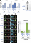
Knockdown of RPL5 or RPL11 retains nucleolar RNA by preventing its export. (A) Knockdown of RPL5 or RPL11 retained RNA content in the nucleolus. The total RNA was isolated from the isolated nucleoli of MCF-7 cells transfected with the indicated siRNAs for 48 h. The total RNA (left) and 28S rRNA (right) levels were quantified by spectrophotometry and RT–qPCR, respectively. The level of the siCont-treated cells was normalized to 1.0. (B) Downregulation of MYBBP1A, RPL5, or RPL11 did not affect the transcription levels of pre-rRNA that had been reduced by TIF-IA knockdown. Nuclear run-on assays were performed to measure the transcription levels of pre-rRNA in MCF-7 cells transfected with the indicated siRNAs for 48 h. Transcription from the rRNA gene was measured by hybridization of in vitro-synthesized 32P-labelled run-on transcripts to immobilized plasmids containing no insert (Control) or cDNA corresponding to 28S rRNA (pre-rRNA). The assays were performed in duplicate. (C) Knockdown of RPL5 or RPL11 prevents nucleolar RNA export from the nucleolus. (Left) MCF-7 cells were transfected with the indicated siRNAs for 48 h. The RNA polymerase II was inhibited by α-amanitin and the newly synthesized rRNA in the cells was pulse labelled with BrUTP. These cells were chased in the BrUTP-free medium for 0 or 60 min, fixed, and stained with anti-BrdU (green), anti-NPM (red) antibodies, or DAPI (blue). (Right) The percentage of cells that showed BrUTP staining in the nucleoplasm at 0 or 60 min after chase. Values are given as the mean±s.d. for triplicate experiments.
Figure 7
Figure 7
Proposed model for the role of RNA content on p53 acetylation by MYBBP1A following nucleolar stress. (A) Model for the role of RNA content in the nucleolus on p53 regulation. See the text. (B) Role of RPL5, RPL11, and MYBBP1A on p53 regulation. See the text.

Similar articles

Cited by

References

    1. Andersen JS, Lam YW, Leung AK, Ong SE, Lyon CE, Lamond AI, Mann M (2005) Nucleolar proteome dynamics. Nature 433: 77–83 - PubMed
    1. Appella E, Anderson CW (2001) Post-translational modifications and activation of p53 by genotoxic stresses. Eur J Biochem 268: 2764–2772 - PubMed
    1. Arva NC, Gopen TR, Talbott KE, Campbell LE, Chicas A, White DE, Bond GL, Levine AJ, Bargonetti J (2005) A chromatin-associated and transcriptionally inactive p53-Mdm2 complex occurs in mdm2 SNP309 homozygous cells. J Biol Chem 280: 26776–26787 - PubMed
    1. Ashcroft M, Kubbutat MH, Vousden KH (1999) Regulation of p53 function and stability by phosphorylation. Mol Cell Biol 19: 1751–1758 - PMC - PubMed
    1. Ashcroft M, Taya Y, Vousden KH (2000) Stress signals utilize multiple pathways to stabilize p53. Mol Cell Biol 20: 3224–3233 - PMC - PubMed

Publication types