
DICER1 deficit induces Alu RNA toxicity in age-related macular degeneration

Hiroki Kaneko et al. Nature. .

Abstract

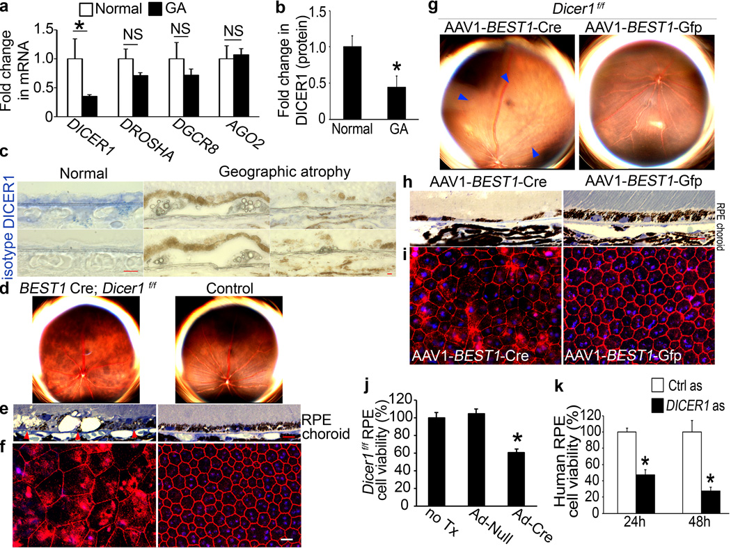
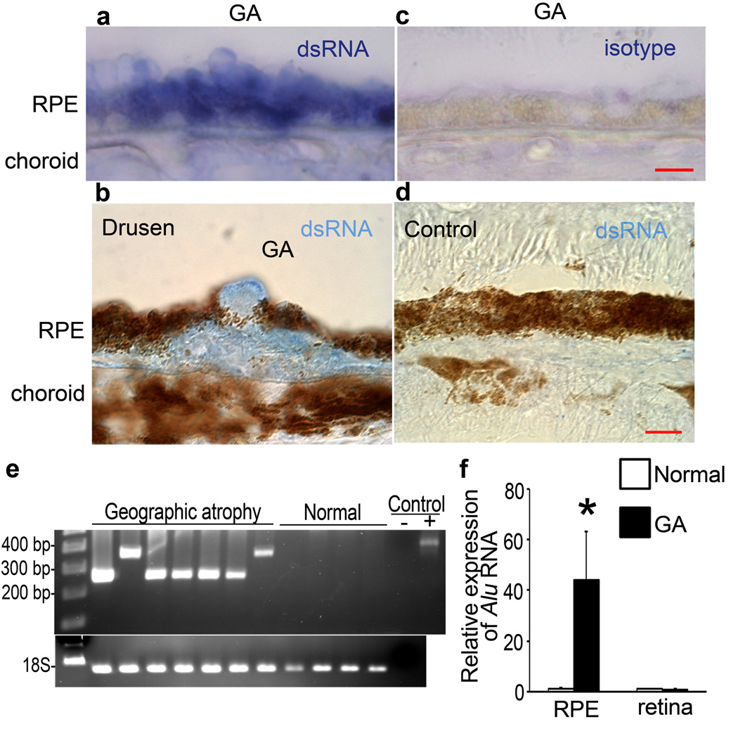
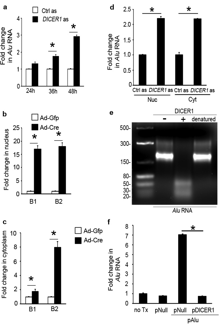
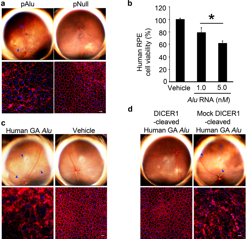
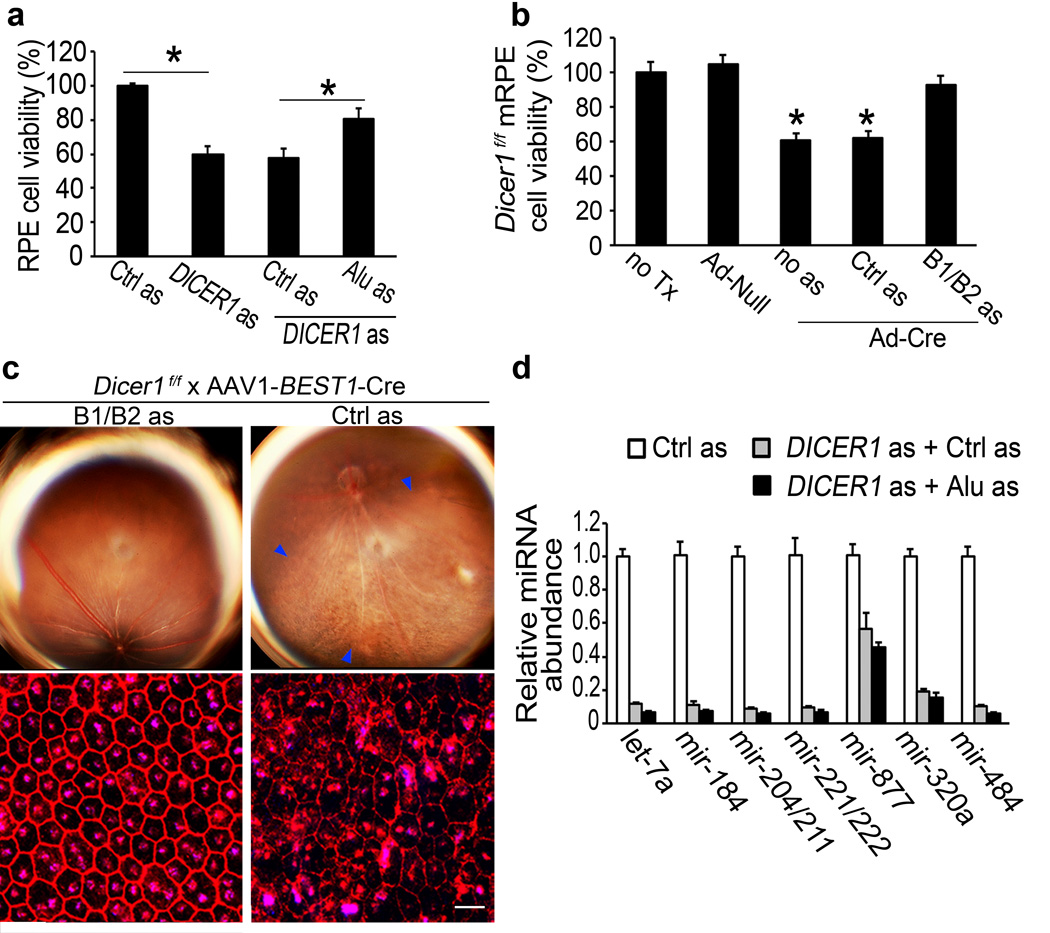
Geographic atrophy (GA), an untreatable advanced form of age-related macular degeneration, results from retinal pigmented epithelium (RPE) cell degeneration. Here we show that the microRNA (miRNA)-processing enzyme DICER1 is reduced in the RPE of humans with GA, and that conditional ablation of Dicer1, but not seven other miRNA-processing enzymes, induces RPE degeneration in mice. DICER1 knockdown induces accumulation of Alu RNA in human RPE cells and Alu-like B1 and B2 RNAs in mouse RPE. Alu RNA is increased in the RPE of humans with GA, and this pathogenic RNA induces human RPE cytotoxicity and RPE degeneration in mice. Antisense oligonucleotides targeting Alu/B1/B2 RNAs prevent DICER1 depletion-induced RPE degeneration despite global miRNA downregulation. DICER1 degrades Alu RNA, and this digested Alu RNA cannot induce RPE degeneration in mice. These findings reveal a miRNA-independent cell survival function for DICER1 involving retrotransposon transcript degradation, show that Alu RNA can directly cause human pathology, and identify new targets for a major cause of blindness.


Figures

Figure 1
Figure 1. DICER1 deficit in GA induces RPE degeneration
a, DICER1 less abundant in RPE of human eyes with GA (n=10) compared to control RPE (n=11). P = 0.004 by Mann Whitney U test. DROSHA, DGCR8, and EIF2C2 (encoding AGO2) abundance not significantly different (P > 0.11 by Mann Whitney U test). n=10–11. b, DICER1 quantification, assessed by Western blotting (Supplementary Fig. 1), lower in human GA RPE (n=4) compared to control RPE (n=4). P = 0.003 by Student t test. c, Reduced DICER1 (blue) in human GA RPE compared to control eyes. d, e, Fundus photographs (d) and toluidine-blue-stained sections (e) show RPE degeneration in BEST1 Cre; Dicer1f/f mice but not controls. Arrowheads point to basal surface of RPE. f, Flatmounts stained for zonula occludens-1 (ZO-1; red) show RPE disruption in BEST1 Cre; Dicerf/f mice compared to controls. g, h, Fundus photographs (g) and toluidine blue-stained sections (h) show RPE (g, h) and photoreceptor (h) degeneration in Dicer1f/f mice following subretinal injection of AAV1-BEST1-Cre but not AAV1-BEST1-GFP. i, Flatmounts show Dicer1f/f mouse RPE degeneration following subretinal injection of AAV1-BEST1-Cre but not AAV1-BEST1-GFP. Nuclei stained blue with Hoechst 33342. Representative images shown. n=16–32 (d–f); 10–12 (g–i). Scale bars, (c,e,h), 10 µm; (f,i) 20 µm. j, Adenoviral vector coding for Cre recombinase (Ad-Cre) treatment reduces Dicer1f/f mouse RPE cell viability compared to Ad-Null or untreated (no Tx) cells. k, DICER1 antisense (as) reduces human RPE cell viability compared to control antisense (Ctrl as)-treated cells. n=6–8. All error bars indicate mean±s.e.m.
Figure 2
Figure 2. Alu RNA accumulation in GA triggered by DICER reduction
a, b, dsRNA immunolocalized (blue) in RPE (a, b) and sub-RPE deposits (drusen; b) in human GA. c, d, No staining with isotype antibody in GA RPE (c) and with anti-dsRNA antibody in control eye (d). Scale bars, (a–d), 10 µm. n=10 (a–d) e, PCR amplification of immunoprecipitated dsRNA yielded amplicons with homology to Alu in GA RPE but not normal RPE. Water control (−) showed no amplification and recombinant dsRNA (+) showed predicted amplicon. f, Increased Alu RNA in GA RPE compared to control (n=7). P < 0.05 by Student t test. No significant difference in Alu RNA in neural retina. Values normalized to abundance in normal eyes.
Figure 3
Figure 3. DICER1 degrades Alu RNA
a, DICER1 antisense (as) increased Alu RNA in human RPE cells. b, c, Ad-Cre, but not Ad-GFP, increased B1 and B2 RNAs in Dicer1f/f mouse RPE cells in nucleus (b) and cytoplasm (c). d, DICER1 as upregulated Alu RNA in human RPE cell nucleus (Nuc) and cytoplasm (Cyt). e, Agarose gel electrophoresis shows recombinant DICER1 (+), but not heat denatured DICER1, degrades Alu RNA isolated and cloned from human GA RPE. Image representative of 6 experiments. f, Alu RNA in human RPE cells upregulated by plasmid coding for Alu (pAlu) vs. pNull or no treatment (no Tx) at 24 h reduced by pDICER1. * P < 0.05. n=4–8 (a–d, f). Values normalized to control as-treated (for Alu) or Ad-GFP-infected cells (for B elements).
Figure 4
Figure 4. DICER1 protects RPE cells from Alu RNA cytotoxicity
a, Subretinal pAlu, but not pNull, induced wild-type mouse RPE degeneration (fundus photographs, top row; ZO-1 stained (red) flatmounts, bottom row). b, Alu RNA induced human RPE cytotoxicity. Values normalized to pNull or vehicle. * P < 0.05 by Student t test. n=4–6. c, Subretinal Alu RNA isolated and cloned from human GA RPE induced wild-type mouse RPE degeneration. d, Subretinal injection of this Alu RNA, when cleaved by DICER1, did not induce wild-type mouse RPE degeneration (fundus photographs, top row; flatmounts, bottom row) in contrast to mock-cleaved Alu RNA. Degeneration outlined by blue arrowheads (a,c,d). Scale bars (20 µm). n=10–15.
Figure 5
Figure 5. DICER1 dyregulation induces RPE cell death via Alu RNA accumulation
a, Human RPE cytotoxicity induced by DICER1 as rescued by Alu RNA as. Values normalized or compared to control (Ctrl) as. b, Ad-Cre but not Ad-Null induced Dicer1f/f mouse RPE cytotoxicity. B1/B2 RNA as, but not control (Ctrl) as, rescued viability. Values normalized to untreated cells (no Tx). * P < 0.05 by Student t test. n=4–6 (a,b). c, Subretinal AAV-BEST1-Cre induced RPE degeneration (blue arrowheads in fundus photograph, top row; ZO-1 stained (red) flatmounts, bottom row) in Dicer1f/f mice 20 days after injection was inhibited by subretinal cholesterol-conjugated B1/B2 as, but not cholesterol-conjugated Ctrl as, 10 days after AAV-BEST1-Cre injection. Values normalized to Ctrl as-treatment. n=8. Scale bar, 20 µm. d, DICER1 as induced global miRNA expression deficits in human RPE cells compared to Ctrl as. No significant difference in miRNA abundance between Alu as and Ctrl as-treated DICER1 depleted cells. n=3.

Comment in

  • Vision: Dicer leaps into view.
    Meister G. Meister G. Nature. 2011 Mar 17;471(7338):308-9. doi: 10.1038/471308a. Nature. 2011. PMID: 21412326 No abstract available.

Similar articles

Cited by

References

    1. Kleinman ME, et al. Sequence- and target-independent angiogenesis suppression by siRNA via TLR3. Nature. 2008;452:591–597. - PMC - PubMed
    1. Takeda A, et al. CCR3 is a target for age-related macular degeneration diagnosis and therapy. Nature. 2009;460:225–230. - PMC - PubMed
    1. Ferrara N. Vascular endothelial growth factor and age-related macular degeneration: from basic science to therapy. Nat Med. 2010;16:1107–1111. - PubMed
    1. Ambati J, Ambati BK, Yoo SH, Ianchulev S, Adamis AP. Age-related macular degeneration: etiology, pathogenesis, and therapeutic strategies. Surv Ophthalmol. 2003;48:257–293. - PubMed
    1. Bernstein E, Caudy AA, Hammond SM, Hannon GJ. Role for a bidentate ribonuclease in the initiation step of RNA interference. Nature. 2001;409:363–366. - PubMed

Publication types

MeSH terms

Associated data