. 2011 Mar;14(3):357-65.
doi: 10.1038/nn.2753. Epub 2011 Feb 6.

The neural substrates of rapid-onset Dystonia-Parkinsonism

Affiliations

The neural substrates of rapid-onset Dystonia-Parkinsonism

D Paola Calderon et al. Nat Neurosci. 2011 Mar.

Abstract

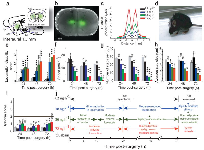
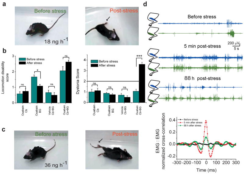
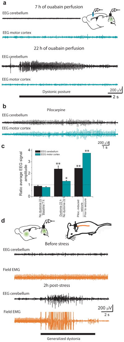
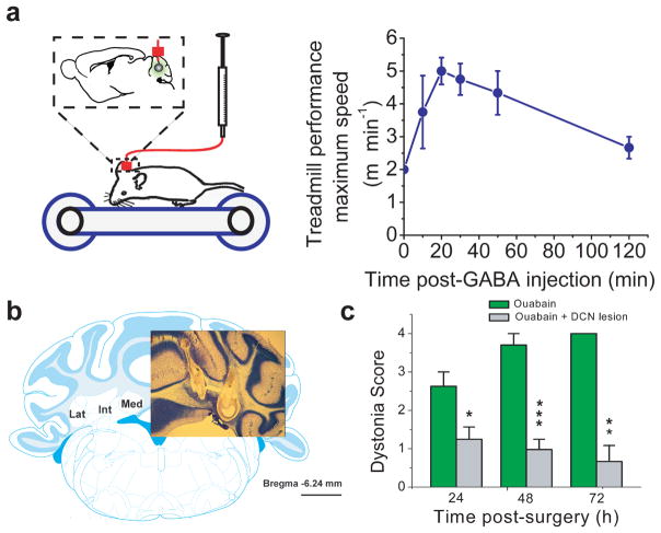
Although dystonias are a common group of movement disorders, the mechanisms by which brain dysfunction results in dystonia are not understood. Rapid-onset Dystonia-Parkinsonism (RDP) is a hereditary dystonia caused by mutations in the ATP1A3 gene. Affected individuals can be free of symptoms for years, but rapidly develop persistent dystonia and Parkinsonism-like symptoms after a stressful experience. Using a mouse model, we found that an adverse interaction between the cerebellum and basal ganglia can account for the symptoms of these individuals. The primary instigator of dystonia was the cerebellum, whose aberrant activity altered basal ganglia function, which in turn caused dystonia. This adverse interaction between the cerebellum and basal ganglia was mediated through a di-synaptic thalamic pathway that, when severed, alleviated dystonia. Our results provide a unifying hypothesis for the involvement of cerebellum and basal ganglia in the generation of dystonia and suggest therapeutic strategies for the treatment of RDP.


Figures

Figure 1
Figure 1. Chronic partial blockade of basal ganglia sodium pumps induces Parkinsonism-like symptoms
(a) Sagittal and coronal schematics of the basal ganglia showing the anatomical structures targeted by chronic bilateral perfusion of ouabain. Use of bodipy-FL-ouabain, a fluorescent analogue of ouabain, allowed determination of the extent of its diffusion in vivo. (b) Merge of the bright field picture of a coronal section of the basal ganglia with the fluorescence intensity profile obtained after perfusion of 18 ng/h bodipy-FL-ouabain for 72 h. (c) The concentration of ouabain in the tissue was estimated by quantitative fluorescence microscopy using the fluorescence emitted by Bodipy-FL-ouabain. The highest concentration of ouabain was obtained close to the canula and dropped monotonically as a function of lateral distance. The color key legend applies to all panels in this figure. (d) Hunched posture, rigidity and akinesia in a mouse after 72 h chronic perfusion of 36 ng/h ouabain. (e–h) Effects of chronic perfusion of 0–72 ng/h ouabain into the basal ganglia on overall locomotion (e), speed (f), average number of steps (g) and step size (h) as a function of time after start of ouabain perfusion. (mean±s.e.m; n= 4 for vehicle; 4 for 7.2 ng/h ouabain; 13 for 18 ng/h; 11 for 36 ng/h; and 3 for 72 ng/h). (i) Assessment of the impact of bilateral perfusion of ouabain into the basal ganglia on motor function using a dystonia rating scale for the same animals described above. With the scale used, only a score of 2 or more denotes dystonia. mean±s.e.m. (j) Summary of symptoms associated with chronic bilateral perfusion of ouabain into the basal ganglia for 24, 48 and 72 hours.
Figure 2
Figure 2. Chronic partial blockade of cerebellar sodium pumps results in ataxia and dystonic-like postures
(a) Coronal schematic of the cerebellum showing the regions affected by chronic perfusion of ouabain. (b) Merge of the bright field picture of a coronal section of the cerebellum with the fluorescence intensity profile obtained after perfusion of 18 ng/h bodipy-FL-ouabain for 18 h. (c) Estimated concentration of ouabain in the cerebellum as a function of lateral distance from the center of the canula with different concentrations of perfused ouabain. (d) Dystonic postures in a mouse caused by perfusion of 36 ng/h ouabain into the cerebellum for 18 hours. Arrows point to the commonly observed hyperextensions of anterior and posterior limbs. (e) Effects of chronic perfusion of 0–72 ng/h ouabain into the cerebellum on locomotor activity in mice at 24, 48 and 72 hours. The decreases in locomotion reflected in the locomotion disability scores were a reflection of the ataxia and dystonia in these mice and not akinesia and rigidity as seen when ouabain was perfused into the basal ganglia. mean±s.e.m. (f) Severity of ouabain-induced dystonia in mice chronically perfused with 0–72 ng/h ouabain into the cerebellum at 4, 12, 24, 48 and 72h. mean±s.e.m. (g) Symptoms associated with chronic perfusion of ouabain into the cerebellum.
Figure 3
Figure 3. Stress-induced dystonia in mice requires interaction between cerebellar and basal ganglia motor control loops
(a) Concomitant perfusion of 18 ng/h ouabain into the cerebellum and basal ganglia for 24 h produces reduced locomotion, and mild gait disturbance (before stress). Immediately after exposing the animal to severe stress the mouse developed persistent dystonic postures. Arrow points to hyperextension of posterior limb. (b) The effect of stress on locomotion and dystonia in mice chronically perfused with 18 ng/h ouabain only in the cerebellum or basal ganglia, or concomitantly in both structures. Stress induces dystonia only in animals in which both the cerebellum and basal ganglia were concomitantly perfused with ouabain. (c) EMGs recorded from agonist and antagonist anterior cranial tibial and gastrocnemius muscles in a mouse in which the cerebellum and basal ganglia were concurrently perfused with 18 ng/h ouabain for 24 h. Before exposure to stress, the mouse showed reduced locomotion but rarely co-contraction of the two muscles. Five minutes post-stress persistent co-contraction of the two muscles could be seen for several seconds. These co-contractions reduced in frequency and intensity, but nonetheless were notable even 3 days later. The graph at the bottom shows normalized cross-correlation of the two EMG signals (mean±s.e.m.). Significant cross-correlation in the activity of the two muscles was observed after, but not before stress. Note that the EMG traces used for analysis corresponded to times at which significant activity in at least one muscle was detected. (d) More severe symptoms in the form of reduced locomotion, and gait disturbance/mild ataxia in a mouse concomitantly perfused with 36 ng/h ouabain into the cerebellum and basal ganglia for 5 h (before stress). Subjecting the mouse to the stress paradigm resulted in generalized dystonia (post stress) including the distortion of the lower jaw.
Figure 4
Figure 4. Dystonic postures correlate with abnormal cerebellar activity
(a) EEG recordings from the motor cortex and the cerebellum of a mouse whose cerebellum was chronically perfused with 36 ng/h ouabain. The first pair of traces (7 h perfusion) was obtained before any noticeable dystonic postures. Neither the cerebellar EEG (black) nor the motor cortex EEG (cyan) show abnormal activity. The second pair of traces (22 h perfusion) was recorded concurrent with a dystonic posture in the mouse and shows an abnormal cerebellar EEG signal whereas the motor cortex EEG was unremarkable. (b) Abnormal electrical activity in both the cerebellum and the motor cortex of the same mouse during status epilepticus induced by 300 mg/kg pilocarpine. (c) The ratios of average cerebellar and motor cortex EEG signal amplitude under various conditions. The average cerebellar EEG signal amplitude was about 2.5-fold larger when the animal manifested dystonic postures compared to time periods straddling dystonia. At exactly the same time periods, the corresponding change in the average motor cortex EEG signal amplitude was much smaller. Both the cerebellar and motor cortex EEG signal amplitudes increased substantially during pilocarpine-induced seizures. mean±s.e.m. (d) EEG activity in the cerebellum (black) and field EMG signals from the back muscles (orange) were recorded in a mouse in which the cerebellum and basal ganglia were concomitantly perfused with 36 ng/h ouabain. The low impedance of the electrode used for field EMG recording permitted the detection of concerted activity of large group of muscle fibers as an accurate marker of the episodes of generalized dystonia. Prior to stress, the mouse showed mild ataxia/gait disturbance but no dystonic postures. The heart rate is noted in the field EMG with no apparent signs of abnormal activity. The cerebellar EEG signal also did not show any abnormal activity. Exposure of the same mouse to stress (the pair of traces labeled as 2 h post-stress) precipitated repeated dystonic postures, the timings of which were reflected in the field EMG signal. The cerebellar EEG showed abnormal hyperactivity of the cerebellum concurrent with the dystonic postures. In this instance the episode of generalized dystonia partly dislodged the field EMG wire which reduced the amplitude of the EKG to within noise levels.
Figure 5
Figure 5. Reducing aberrant cerebellar activity or silencing cerebellar output lessons dystonia
(a) Mice whose cerebella were chronically perfused with 36 ng/h ouabain showed clear signs of dystonia and were unable to walk on a treadmill at its lowest speed setting of 2 m/min. Acute injection of GABA into their cerebellum using the auxiliary port of the same canula used for ouabain perfusion reduced the severity of their dystonic postures such that, on average, the mice could walk on the treadmill at a pace of 5 m/min. (n=4; mean±s.e.m.) (b) Deep cerebellar nuclei (DCN) were electrically lesioned in both cerebellar hemispheres at either at one or two sites (the two site lesion is shown here). Comparable data was obtained with both approaches. Scale bar corresponds to 1 mm. (c) Chronic perfusion of 36ng/h ouabain into the cerebellum of DCN-lesioned mice did not produce dystonia. (n=7; mean±s.e.m.)
Figure 6
Figure 6. Severing the link between the cerebellum and basal ganglia alleviates dystonia
(a) Using bilateral electrical lesions, the CL nucleus of the thalamus was selectively ablated. The photograph on the left shows Nissl stain of one such lesion, and the schematic on right shows the relevant brain structures with the lesion area marked in red. The scale bar corresponds to 1 mm. Legends correspond to: GP: Globus Palidus, CPu; Caudate/Putaman, and the following nuclei of the thalamus: CM: centrolmedian, CL: centrolateral, VL: ventral lateral, VP: ventral posterior, VM: ventral medial, and Pc: Paracentral. (b) The consequences of CL lesions (or sham operations noted as “No CL lesion”) on the motor symptoms associated with chronic perfusion of 36 ng/h ouabain into the cerebellum of mice was determined by assessing their locomotion and dystonia scores. Lesioning the CL significantly reduced ouabain-induced motor dysfunction and prevented generation of dystonia.

Comment in

Similar articles

Cited by

References

    1. Stacy MA. Handbook of Dystonia. Informa Health care; New York: 2006.
    1. Breakefield XO, et al. The pathophysiological basis of dystonias. Nat Rev Neurosci. 2008;9:222–234. - PubMed
    1. Muller U. The monogenic primary dystonias. Brain. 2009;132:2005–2025. - PubMed
    1. De Carvalho AP, et al. Mutations in the Na+/K+-ATPase alpha3 gene ATP1A3 are associated with rapid-onset dystonia parkinsonism. Neuron. 2004;43:169–175. - PubMed
    1. Brashear A, et al. The phenotypic spectrum of rapid-onset dystonia-parkinsonism (RDP) and mutations in the ATP1A3 gene. Brain. 2007;130:828–835. - PubMed

Publication types

MeSH terms

Supplementary concepts