. 2011 Jan 26;6(1):e14591.
doi: 10.1371/journal.pone.0014591.

Proteasome inhibition represses unfolded protein response and Nox4, sensitizing vascular cells to endoplasmic reticulum stress-induced death

Affiliations

Proteasome inhibition represses unfolded protein response and Nox4, sensitizing vascular cells to endoplasmic reticulum stress-induced death

Angélica M Amanso et al. PLoS One. .

Abstract

Background: Endoplasmic reticulum (ER) stress has pathophysiological relevance in vascular diseases and merges with proteasome function. Proteasome inhibition induces cell stress and may have therapeutic implications. However, whether proteasome inhibition potentiates ER stress-induced apoptosis and the possible mechanisms involved in this process are unclear.

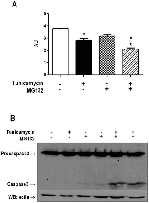
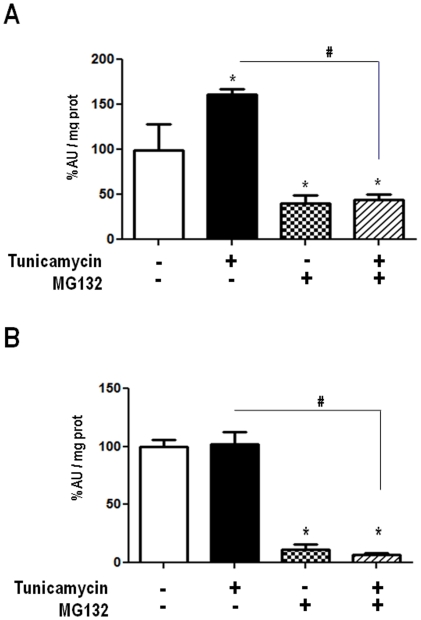
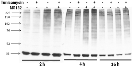
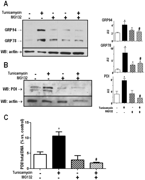
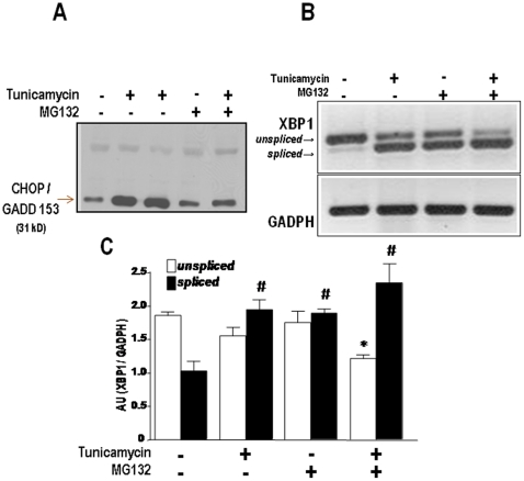
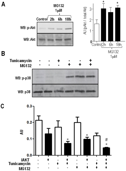
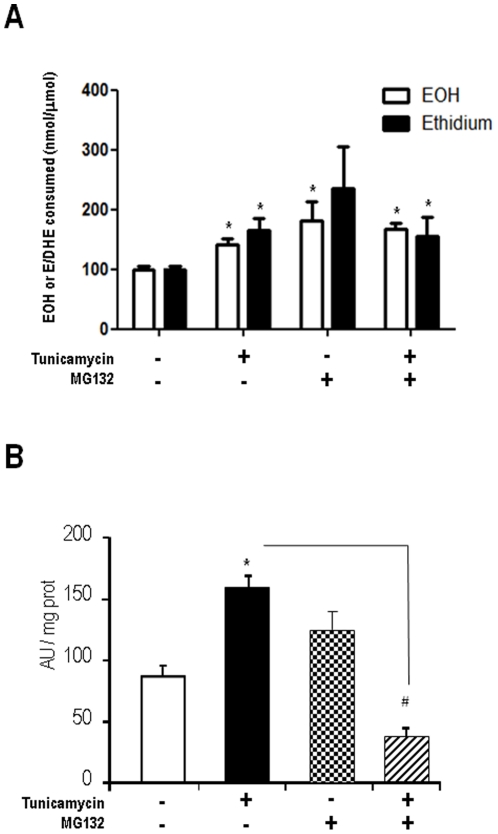
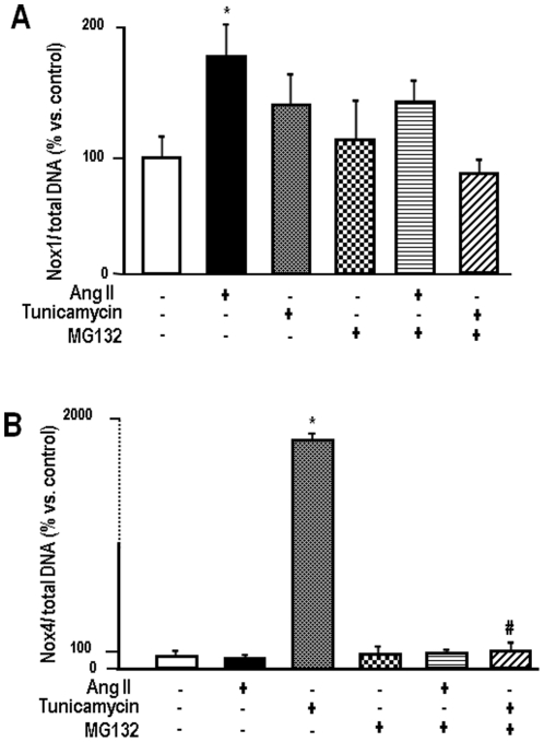
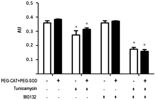
Methodology/principal findings: Here we show that proteasome inhibition with MG132, per se at non-lethal levels, sensitized vascular smooth muscle cells to caspase-3 activation and cell death during ER stress induced by tunicamycin (Tn). This effect was accompanied by suppression of both proadaptive (KDEL chaperones) and proapoptotic (CHOP/GADD153) unfolded protein response markers, although, intriguingly, the splicing of XBP1 was markedly enhanced and sustained. In parallel, proteasome inhibition completely prevented ER stress-induced increase in NADPH oxidase activity, as well as increases in Nox4 isoform and protein disulfide isomerase mRNA expression. Increased Akt phosphorylation due to proteasome inhibition partially offset the proapoptotic effect of Tn or MG132. Although proteasome inhibition enhanced oxidative stress, reactive oxygen species scavenging had no net effect on sensitization to Tn or MG132-induced cell death.

Conclusion/relevance: These data indicate unfolded protein response-independent pathways whereby proteasome inhibition sensitizes vascular smooth muscle to ER stress-mediated cell death. This may be relevant to understand the therapeutic potential of such compounds in vascular disease associated with increased neointimal hyperplasia.


Conflict of interest statement

Competing Interests: The authors have declared that no competing interests exist.

Figures

Figure 1
Figure 1. Proteasome inhibition sensitizes VSMC to death due to ER stress.
(A) Representative graph of VSMC cell viability by MTT assay. VSMC were incubated with tunicamycin (Tn) (5 µg/mL) or/and MG132 (1 µM) for 16 h, followed by MTT assays. (B) Similar to A, VSMC were incubated with Tn (5 µg/mL) or/and MG132 (1 µM) for 16 hours. Total cell homogenates were submitted to western analysis with anti-caspase-3 antibody. Data are mean ± SD of 3 independent experiments.*P<0.05 vs. Control. #P<0.05 vs.Tn.
Figure 2
Figure 2. Activity of 20S proteasome in VSMC at control condition or after incubation with AngII (200 nM) or Tn (5 µg/mL), in the absence or presence of MG132 (1 µM) during 4 (A) or 16 (B) hours.
Cell lysates were incubated with probe AMC (LLVY-AMC) in the presence of SDS and fluorescence release followed over 15–30 min (excitation 355 nm, emission 460 nm). Data are mean ± SD of 4 or more independent experiments.*P<0.05 vs. Control. #P<0.05 vs. Tn.
Figure 3
Figure 3. Total polyubiquitinated protein levels after Tn, MG132 or their combination, at 2, 4 or 16 h of incubation with VSMC.
Western analysis of VSMC lysates was performed in 8% polyacrilamide gels and probed with anti-ubiquitin antibody. Representative of at least 3 experiments per group.
Figure 4
Figure 4. Down-regulation of UPR signaling by MG132.
VSMC were incubated with Tn (5 µg/ml), MG132 (1 µM) or their combination for 16 h. Total cell homogenates were submitted to western analysis with anti-KDEL (A) or anti-PDI antibodies (B). Graphs to the right are corresponding densitometric measurements of blots shown in (A) and (B) for at least 3 independent experiments; (C) Analysis of PDI mRNA by real-time PCR. VSMC were incubated with vehicle or Tn (5 µg/mL) in the absence or presence of MG132 (1 µM) for 16 h; n = 3. Data are mean ± SD. *P<0.05 vs. Control. #P<0.05 vs.Tn.
Figure 5
Figure 5. Effects of proteasome inhibition on nuclear CHOP/GADD153 expression and XBP1 mRNA splicing.
(A) Representative western analysis of CHOP/GADD153 protein expression in nuclear extracts of VSMC incubated with Tn (5 µg/ml) or/and MG132 (1 µM) for 16 h. (B) Agarose gel depicting the amplified PCR products corresponding to spliced or unspliced forms of XBP1 mRNA, obtained from VSMC submitted to the same conditions as in (A). (C) Graph depicting densitometric analysis of data from (B). Data are mean ± SD of 3 independent experiments. *P<0.05 vs. unspliced control. #P<0.05 vs. spliced control.
Figure 6
Figure 6. Akt and p38 phosphorylation by MG132 in VSMC.
(A) Representative immunoblotting depicting phosphorylated and total Akt expression at baseline or after incubation with MG132 (1 µM) for 2, 6 or 16 h. Graph to the right depicts quantitative densitometric analysis of Akt expression from 3 blots similar to (A). (B) Representative immunoblotting depicting phosphorylated and total p38MAPK expression at baseline or after incubation with angiotensin II, tunicamycin, MG132 (1 µM, 16 h) or their combination; representative of n = 3. (C) Graph summarizing the effect of Akt inhibition on enhancement of VSMC death. VSMC were incubated with Tn (5 µg/mL) or/and MG132 (1 µM) for 16 h in the absence or presence of Akt inhibitor (A6730 Sigma-Aldrich), followed by MTT assays. Data are mean ± SD of 3 independent experiments.*P<0.05 vs. Control. #P<0.05 vs.Tn.
Figure 7
Figure 7. Proteasome inhibition induces ROS production and disrupts ER stress-induced NADPH oxidase up-regulation.
(A) ROS production in VSMC incubated for 4 h with Tn (5 µg/mL) or/and MG132 (1 µM), assessed through HPLC analysis of DHE oxidation products (50 µM, 30 min incubation), as described in Methods. Results depict levels of 2-hydroxyethidium (EOH) or ethidium (E) products. (B) NADPH oxidase activity measured in membrane-enriched homogenates from VSMC incubated for 4 h with Tn or MG132. Activity was measured with DHE technique, analogous to (A), as described in Methods. Data are mean ± SD of 3 independent experiments.*P<0.05 vs. Control. #P<0.05 vs.Tn.
Figure 8
Figure 8. Proteasome inhibition strongly inhibits ER stress-induced Nox4 expression.
(A) Real-time PCR analysis of Nox1 mRNA levels. VSMC were incubated with AngII (200 nM) or Tn (5 µg/mL) in the absence or presence of MG132 (1 µM) for 16 h; (B) Similar to (A), with analysis of Nox4 mRNA levels. In both (A) and (B), data are expressed as the ratio of Nox expression/total DNA expression in the same sample. Data are mean ± SD of 5 independent experiments.*P<0.05 vs. Control. #P<0.05 vs. Tn.
Figure 9
Figure 9. Effects of ROS scavenging in VSMC viability after ER stress or/and proteasome inhibition.
VSMC were incubated with Tn (5 µg/mL) or/and MG132 (1 µM) for 16 h in the absence or presence of PEG-Cat (200 U/ml) plus PEG-SOD (25 U/ml). Data are mean ± SD of 3 independent experiments.*P<0.05 vs. Control. #P<0.05 vs.Tn.

References

    1. Zhang K, Kaufman RJ. From endoplasmic-reticulum stress to the inflammatory response. Nature. 2008;454:455–462. - PMC - PubMed
    1. Ron D, Walter P. Signal integration in the endoplasmic reticulum unfolded protein response. Nat Rev Mol Cell Biol. 2007;8:519–529. - PubMed
    1. Marciniak SJ, Ron D. Endoplasmic reticulum stress signaling in disease. Physiol Rev. 2006;86:1133–1149. - PubMed
    1. Rutkowski DT, Kaufman RJ. That which does not kill me makes me stronger: adapting to chronic ER stress. Trends Biochem Sci. 2007;32:469–476. - PubMed
    1. Santos CX, Tanaka LY, Wosniak J, Laurindo FR. Mechanisms and implications of reactive oxygen species generation during the unfolded protein response: roles of endoplasmic reticulum oxidoreductases, mitochondrial electron transport, and NADPH oxidase. Antioxid Redox Signal. 2009;11:2409–2427. - PubMed

Publication types