. 2011 Jan 15:10:2.
doi: 10.4103/1477-3163.75723.

Zoledronic acid directly suppresses cell proliferation and induces apoptosis in highly tumorigenic prostate and breast cancers

Affiliations

Zoledronic acid directly suppresses cell proliferation and induces apoptosis in highly tumorigenic prostate and breast cancers

Hussain Almubarak et al. J Carcinog. .

Retraction in

Abstract

Background: Bisphosphonates (BPs) were designed for the prevention of skeletal-related events secondary to bone metastases. The purpose of this study was to show that zoledronic acid (ZA) directly eradicates highly tumorigenic and potentially metastatic cancer cells.

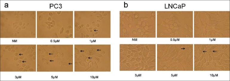
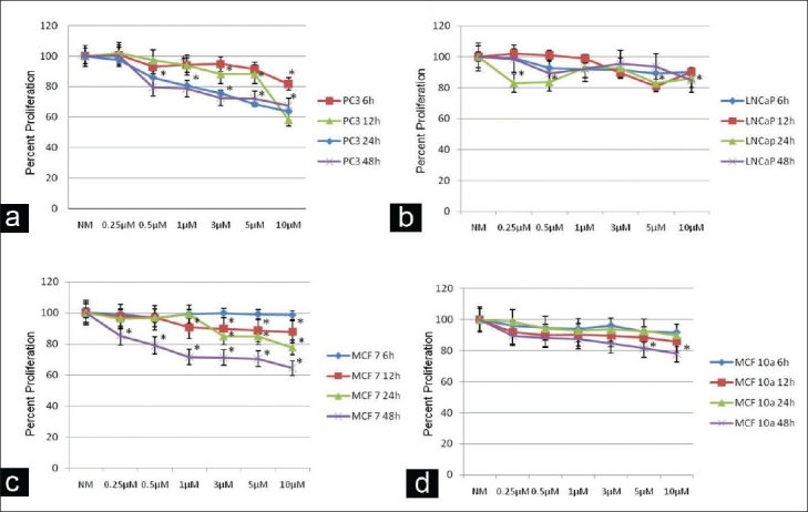
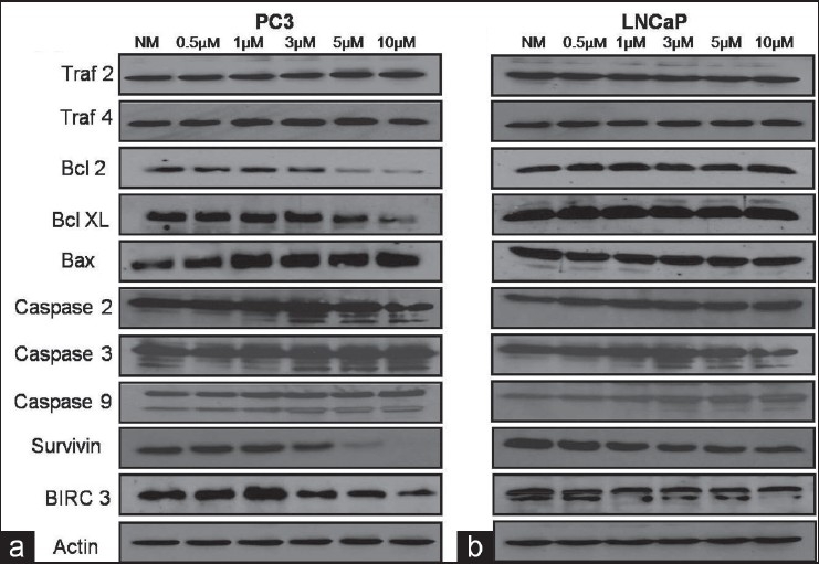
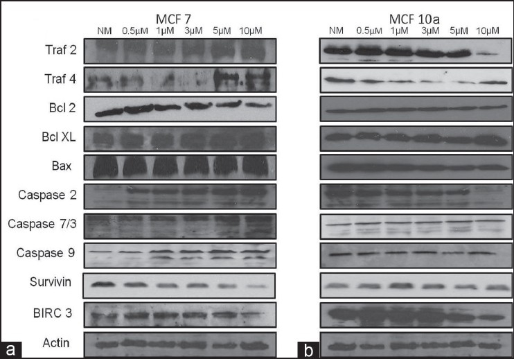
Materials and methods: Human prostate and breast highly tumorigenic (PC3, MCF 7) and low- or non-tumorigenic (LNCaP, MCF 10a) cell lines, respectively, were exposed to different concentrations of ZA (0-10 μM). Reverse transcriptase double quantitative polymerase chain reaction was used for quantitative gene expression analysis. Apoptosis and cell proliferation were determined using microscopic observation and MTS assays. Western blot was used to confirm the translational effects of apoptotic genes on protein expression.

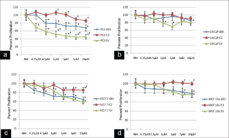
Results: Human prostate and breast highly tumorigenic (PC3, MCF 7) and low- or non-tumorigenic (LNCaP, MCF 10a) cell lines, respectively, showed multiple genes demonstrating differential expressions, including TRAF, TRADD, BCL2, CASPASES and IAP families. Increasing ZA concentrations showed a greater concentration-time response on cell proliferation and apoptosis in the highly tumorigenic cells. These results were confirmed by both reversing and enhancing the effect of ZA on cell proliferation with caspase 3, 7 or survivin siRNA, respectively. Pro-apoptotic proteins bax and caspase 2, 3, 7 and 9 were up-regulated, while the anti-apoptotic proteins bcl2, birc3 and survivin were down-regulated only in the highly tumorigenic cells.

Conclusions: This explains the ability of ZA to inhibit bony metastasis in highly tumorigenic cells compared with the low- or non-tumorigenic cells through a significant decrease in cell proliferation and increase in apoptosis through gene-regulated and translational-mediated down-regulation of survivin coupled with the inhibition of caspase 3 or 7. This has significant implications toward understanding the pharmacophysiology of BPs in metastasis and supports the clinically observed effect of BPs when administered adjunctively with anticancer drugs such as cyclophosphamide/methotrexate/5-fluorouracil, epirubicin in combination with cyclophosphamide or docetaxel, and doxorubicin.

Keywords: Apoptosis; bisphosphonate; cancer; metastasis; zoledronic acid.


Figures

Figure 1
Figure 1
Microscopic apoptosis analysis: (a) microscopic images of apoptosis in human prostate highly tumorigenic PC3 cells using either treatment with normal media with infusion solution (NM-Control) or zoledronic acid (ZA) at 0.5, 1, 3, 5 or 10 μM; showing the rounding, balling up and fragmentation of cells starting at 1 μM, with apoptotic effects seen in a dose-dependent manner, with the greatest effects seen at 5 and 10 μM (arrows) (×400 magnification). (b) Microscopic images of apoptosis in human low tumorigenic LNCaP cells using either treatment with normal media with infusion solution (NM) or ZA at 0.5, 1, 3, 5 or 10 μM; showing no significant difference with increasing ZA concentrations, showing only minimal changes at 5 and 10 μM (arrows). Cells were observed over 24 h, with images taken at 24 h (original magnification, ×400)
Figure 2
Figure 2
Cell proliferation: assessment of human prostate (PC3, LNCaP) and breast (MCF 7, MCF 10a) cancer cell proliferation using MTS assay following 6, 12, 24 or 48 h treatment compared with control cells and normal media with infusion solution (NM). (a, c) Decrease in tumor cell proliferation in a dose- and time-dependent manner in PC3 and MCF 7 cells following exposure to NM, 0.25, 0.5, 1, 3, 5 or 10 µM zoledronic acid (ZA) compared with control cells (NM). (b, d) Minimal decrease in tumor cell proliferation in LNCaP and MCF 10a cells following exposure to NM, 0.25, 0.5, 1, 3, 5 or 10 µM ZA compared with control cells (NM), showing that only high doses and long time periods decreased cell proliferation. Error bars indicate the standard errors of the means. The growth of control cells (NM) was set to 100%. *Statistically significant (P < 0.05) differences compared with control cells
Figure 3
Figure 3
Alteration of cell proliferation: human prostate (PC3, LNCaP) and breast (MCF 7, MCF 10a) cancer cells following 48 h of treatment with normal media with infusion solution (NM) or 0.25, 0.5, 1, 3, 5 or 10 μM zoledronic acid (ZA) or these treatments combined with siRNA against caspase 3, 7 or survivin. (a, c) Decrease in cell proliferation in a dose- and time-dependent manner in PC3 and MCF 7 cells following exposure to 0.25, 0.5, 1, 3, 5 or 10 μM ZA compared with the control cells (NM). Caspase 3 siRNA signifi cantly reversed the effects of ZA on cell proliferation, starting at ZA concentrations of 1 μM in PC3 cells and Caspase 7 siRNA signifi cantly reversed the effects of ZA on cell proliferation, starting at ZA concentrations of 10 μM in MCF 7 cells compared with cells treated with ZA alone. Survivin siRNA showed a significant additional decrease in cell proliferation starting at ZA 1 μM in PC3 cells compared with cells treated with ZA alone. MCF 7 cells showed a decrease in cell proliferation at higher ZA concentrations; however, this was not significant. (b, d) Minimal decrease in tumor cell proliferation in a dose- and time-dependent manner in LNCaP and MCF 10a cells following exposure to 0.25, 0.5, 1, 3, 5 or 10 μM ZA compared with control cells (NM). Results demonstrate that caspase 3 siRNA in LNCaP cells significantly reversed the effects of ZA on cell proliferation at ZA concentrations of 5 and 10 μM. Survivin siRNA showed no significant additional decrease in cell proliferation. The growth of control cells (NM) was set to 100%. *Statistically significant (P < 0.05) differences compared with control cells (NM) for ZA only and control-scrambled siRNA for Caspase 3, 7 and survivin treated. †Statistically significant (P < 0.05) differences following siRNA treatment compared with ZA treatment on a dose-dependent basis
Figure 4
Figure 4
Western blot analysis: assessment of human prostate cancer cells (a) PC3 and (b) LNCaP demonstrating that following 24 h of exposure to normal media with infusion solution (NM) or 0.5, 1, 3, 5 and 10 μM doses of zoledronic acid (ZA), several apoptotic and anti-apoptotic proteins were differentially expressed in the highly tumorigenic PC3 cell line. Several apoptotic proteins were over-expressed, including bax and the increased cleavage of caspase proteins 2, 3 and 9, with increasing dose of ZA. Additionally, several anti-apoptotic proteins were downregulated in PC3 cells, including bcl2, bclxl, survivin and birc3. Surface death domain proteins, traf2 and 4 showed minimal change. Actin was used as the loading control
Figure 5
Figure 5
Western blot analysis: assessment of human breast cancer cells (a) MCF 7 and (b) MCF 10a demonstrating that following 24 h of exposure to normal media with infusion solution (NM) or 0.5, 1, 3, 5 and 10 μM doses of ZA, several apoptotic and anti-apoptotic proteins were differentially expressed in the highly tumorigenic MCF 7 cell line. Several apoptotic proteins were overexpressed, including the increased cleavage of caspase proteins 2, 7 and 9 with increasing dose of ZA. Additionally, several anti-apoptotic proteins were down-regulated in MCF 7 cells, including bcl2, survivin and birc3. Surface death domain protein, traf4 showed up-regulation in MCF 7 cells. Actin was used as the loading control

Similar articles

Cited by

References

    1. Jemal A, Siegel R, Ward E, Hao Y, Xu J, Thun MJ. Cancer statistics, 2009. CA Cancer J Clin. 2009;59:225–49. - PubMed
    1. Drake MT, Clarke BL, Khosla S. Bisphosphonates: mechanism of action and role in clinical practice. Mayo Clin Proc. 2008;83:1032–45. - PMC - PubMed
    1. Lyseng-Williamson KA. Zoledronic acid: A review of its use in breast cancer. Drugs. 2008;68:2661–82. - PubMed
    1. Ross JR, Saunders Y, Edmonds PM, Patel S, Wonderling D, Normand C, et al. A systematic review of the role of bisphosphonates in metastatic disease. Health Technol Assess. 2004;8:1–176. - PubMed
    1. Pavlakis N, Schmidt R, Stockler M. Bisphosphonates for breast cancer. Cochrane Database Syst Rev. 2005;3 CD003474. - PubMed

Publication types