. 2011 Jan 31;6(1):e14600.
doi: 10.1371/journal.pone.0014600.

CDK11(p58) is required for centriole duplication and Plk4 recruitment to mitotic centrosomes

Affiliations

CDK11(p58) is required for centriole duplication and Plk4 recruitment to mitotic centrosomes

Nathalie Franck et al. PLoS One. .

Abstract

Background: CDK11(p58) is a mitotic protein kinase, which has been shown to be required for different mitotic events such as centrosome maturation, chromatid cohesion and cytokinesis.

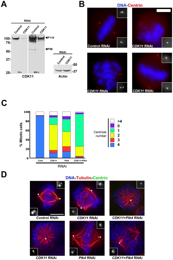
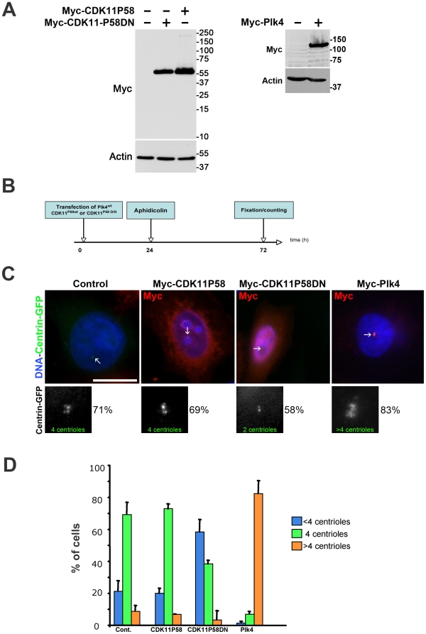
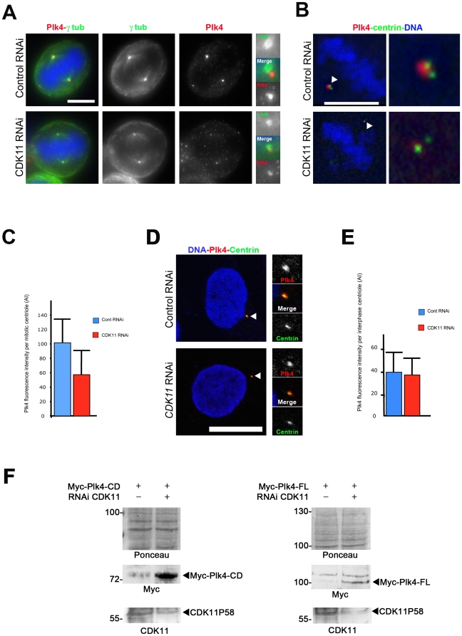
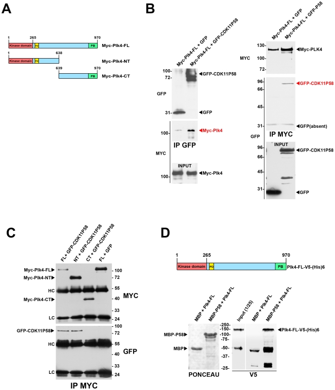
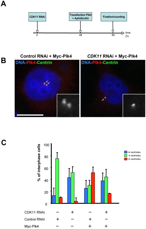
Methodology/principal findings: In addition to these previously described roles, our study shows that CDK11(p58) inhibition induces a failure in the centriole duplication process in different human cell lines. We propose that this effect is mediated by the defective centrosomal recruitment of proteins at the onset of mitosis. Indeed, Plk4 protein kinase and the centrosomal protein Cep192, which are key components of the centriole duplication machinery, showed reduced levels at centrosomes of mitotic CDK11-depleted cells. CDK11(p58), which accumulates only in the vicinity of mitotic centrosomes, directly interacts with the centriole-associated protein kinase Plk4 that regulates centriole number in cells. In addition, we show that centriole from CDK11 defective cells are not able to be over duplicated following Plk4 overexpression.

Conclusion/significance: We thus propose that CDK11 is required for centriole duplication by two non-mutually-exclusive mechanisms. On one hand, the observed duplication defect could be caused indirectly by a failure of the centrosome to fully maturate during mitosis. On the other hand, CDK11(p58) could also directly regulate key centriole components such as Plk4 during mitosis to trigger essential mitotic centriole modifications, required for centriole duplication during subsequent interphase.


Conflict of interest statement

Competing Interests: The authors have declared that no competing interests exist.

Figures

Figure 1
Figure 1. CDK11p58 depletion leads to a reduction in centriole number in mitotic HeLa cells.
Mitotic HeLa cells stably expressing a GFP-tagged centrin were subjected to control or CDK11 RNAi and analysed after 72 hours. A) Western blots showing CDK11p110 and CDK11p58 (with 10 and 600 seconds exposure times respectively) and actin protein levels are shown. The positions of CDK11p110 and CDK11p58 are indicated. B) Mitotic cells stained for DNA (blue) and centrin (red) following control or CDK11 RNAi. Some of the observed defects are shown here. The insets show a 3× magnification view of the centriole region in monochrome. The treatment is displayed at the bottom of each panel. Scale bar is 5µm. C) Quantitative analysis of the centriole distribution following control, CDK11, Plk4 or double RNAi in mitotic cells. See the reduced number of centrioles following CDK11 RNAi (also shown in Table S1). D) Example of the mitotic figures observed in control, CDK11, Plk4 and double (CDK11 and Plk4) siRNA–transfected cells. See also detailed analysed in Table S1. Centrioles are green, microtubules are red and chromosomes are blue. The insets show a 3× magnification view of the centriole region in monochrome (indicated by a white triangle in the merge panels). Bar is 5µm.
Figure 2
Figure 2. Kinase-dead CDK11p58D/N but not wild-type CDK11p58 overexpression leads to a centriole duplication failure in S-phase arrested HeLa cells.
A) Expression of Myc-CDK11p58 and Myc-CDK11p58D/N (left) or Myc-Plk4 proteins following transfection of the appropriate constructs. A Myc and an actin (as a loading control) Western blotting are shown. B) HeLa cells stably expressing GFP-tagged centrin were transfected with either active (CDK11p58) or inactive (CDK11p58DN) CDK11p58 expression constructs. A positive control was also performed using Myc-Plk4. After 24 hours (duration of one cell cycle), the cells were arrested in S phase by aphidicolin treatment for 24 hours and the centrioles were counted after 48 more hours. C) The transfected cells expressing Myc-tagged proteins were visualised using an anti-Myc antibody (red). DNA is blue. The centriole (green), indicated by arrows, (also displayed in monochrome in the lower panels) were counted in control or transfected cells. In cells expressing CDK11p58 as well as in control cells, the vast majority of the cells (∼70%) displayed four centrioles. This percentage dropped to 38.2% when CDK11p58DN was expressed. Note that more than 80% of the Plk4 over expressing cells showed centriole amplification. Scale bar is 10µm. D) Graph (±SD) showing the percentage of cells with <4 centrioles (blue), 4 (green) or >4 (orange).
Figure 3
Figure 3. CDK11 depleted cells show diminished recruitment of the Plk4 protein at the mitotic centrosomes.
A) HeLa cells were transfected with control or CDK11 siRNAs and stained for Plk4 (red and right panels in monochrome) and γ tubulin (green and middle panels in monochrome). In the absence of CDK11p58, Plk4 signal is strongly reduced at the centrosome of mitotic cells. For each cell, one of the two centrosomal regions is enlarged on the left panel. Bar is 5µm. B) Single section of a control or CDK11 siRNA treated cell stained for centrin (green) and Plk4 (red). The insets show a 5× magnification of the spindle pole region that contains 2 centrioles. See the strong reduction of the Plk4 signal in the CDK11–depleted cell compared to the wild-type cell. Bar is 5µm. C) Graph showing the significant difference (p<0,0005) of Plk4 signal intensity (±SD) per mitotic centriole in control (blue) or CDK11 siRNA (red) transfected cells. D) Control (top) or CDK11 (bottom) siRNAs-transfected HeLa interphase cells stained for Plk4 (red), centrin (green). The right panels show a 3× magnification of the centrosomal region. Bar is 10µm. E) Graph showing the Plk4 signal (±SD) intensity per interphase centriole in control (blue) or CDK11 siRNA transfected cells (red). F) Analysis of exogenous Plk4 protein levels in CDK11 knock down cells. Myc-Plk4-CD (Catalytic Domain; amin-acids 1–638, left panels) and Myc-Plk4-FL (Full Length; amino-acids 1–970, right panels) expressing constructs were co-transfected with control or CDK11 siRNAs and the CDK11p58 (bottom panels) and Myc-Plk4 (middle panels) proteins levels were analysed by Western blotting 48 h following transfection. The membrane was stained by Ponceau S (top panels). In four different experiments, exogenous Plk4 proteins are hardly detectable after 48h hours in control cells whereas they are more stable following CDK11 RNAi.
Figure 4
Figure 4. Plk4 and CDK11p58 can interact in vitro and in vivo.
A) Scheme of the different Plk4 constructs used for the co-transfection and immunoprecipitation experiments. Myc-Plk4-FL: Myc-tagged Plk4 full length; Myc-Plk4-NT: Myc-tagged Plk4 N-terminus domain; Myc-Plk4-CT: Myc-tagged Plk4 C-terminus domain. The catalytic domain (CD) is displayed in red, the polo box (PB) in green and the phosphodegron (PD) in yellow. B) GFP or GFP-CDK11p58 were co-transfected with Myc-Plk4 in COS7 cells and the proteins were immunoprecipitated using GFP (left) or Myc antibodies (right). Myc-Plk4 was co-immunoprecipitated with GFP-CDK11p58 but not with GFP (left). GFP-CDK11p58 (but not GFP) was also found in the Myc-Plk4 immunoprecipitates (right). C) Myc-Plk4-FL, Myc-Plk4-NT, Myc-Plk4-CT were co-transfected with GFP and/or GFP-CDK11p58 in COS7 cells. Myc-tagged Plk4 proteins were immunoprecipitated and revealed by Myc (top)(bottom). GFP-CDK11p58 is detected in Myc-Plk4-FL and NT immunoprecipitates whereas it is absent from Myc-Plk4-CT pull down (bottom). HC: immunoglobulin heavy chains; LC: immunoglobulin low chains. D) Maltose Binding proteins (MBP) or MBP-CDK11p58 proteins were immobilised on amylose beads and incubated with recombinant Plk4-V5-(His)6. After washes, the beads (and bound proteins) were boiled in Laemmli buffer and the proteins were analysed following EGPA-SDS by Ponceau S staining to visualise MBP and MBP-CDK11p58 (left). The membrane was then probed by anti-V5 antibody to reveal the bound Plk4 recombinant protein. The input (1/25th) is also shown on the gel. Thus, Plk4 and CDK11p58 can bind directly to each other.
Figure 5
Figure 5. CDK11-depleted cells cannot amplify their centrioles following Plk4 overexpression.
A) Control or CDK11-depleted cells were transfected with Myc-Plk4 construct and their ability to amplify their centrioles was analysed after S phase arrest. B) Example of a control siRNA treated cell (left) over expressing Plk4 (red). See the presence of additional centrin (green) dots around each mother centriole. By contrast, most of Myc-Plk4 over expressing cells can not amplify their centrioles following CDK11 siRNA transfection although the over expressed Plk4 protein kinase is strongly recruited to the centriolar region.The insets shows a 5× magnification of the centrosomal region (centrin staining in displayed in monochrome). Bar is 10µm.

Similar articles

Cited by

References

    1. Kellogg DR, Moritz M, Alberts BM. The centrosome and cellular organization. Annu Rev Biochem. 1994;63:639–674. - PubMed
    1. Nigg EA. Centrosome duplication: of rules and licenses. Trends Cell Biol. 2007;17:215–221. - PubMed
    1. Nigg EA. Centrosome aberrations: cause or consequence of cancer progression? Nat Rev Cancer. 2002;2:815–825. - PubMed
    1. Nigg EA. Origins and consequences of centrosome aberrations in human cancers. Int J Cancer. 2006;119:2717–2723. - PubMed
    1. Delattre M, Leidel S, Wani K, Baumer K, Bamat J, et al. Centriolar SAS-5 is required for centrosome duplication in C. elegans. Nat Cell Biol. 2004;6:656–664. - PubMed

Publication types