. 2011 Jan 31;6(1):e16172.
doi: 10.1371/journal.pone.0016172.

Efficacy of fumaric acid esters in the R6/2 and YAC128 models of Huntington's disease

Affiliations

Efficacy of fumaric acid esters in the R6/2 and YAC128 models of Huntington's disease

Gisa Ellrichmann et al. PLoS One. .

Abstract

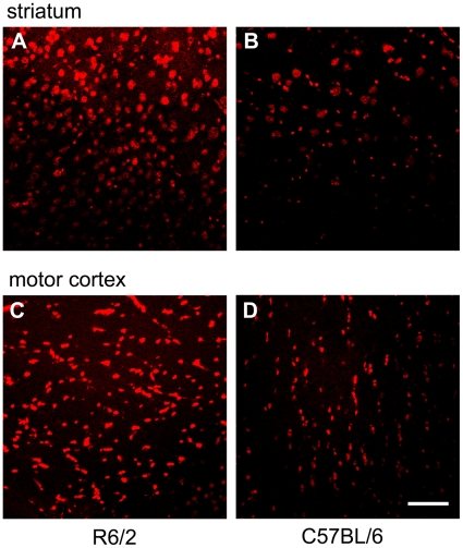
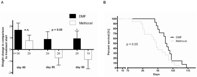
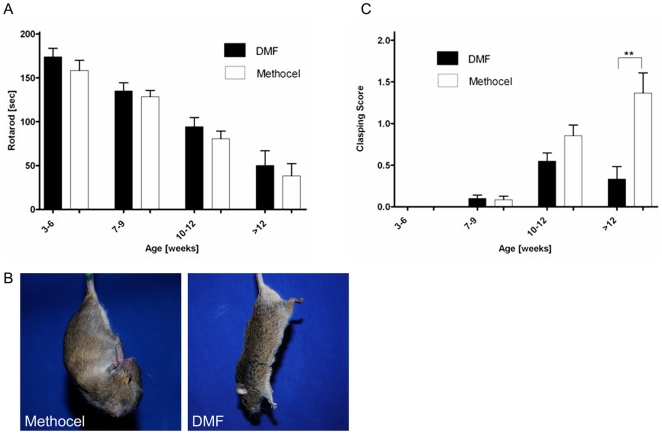
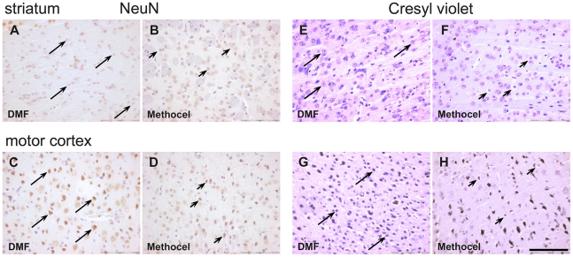
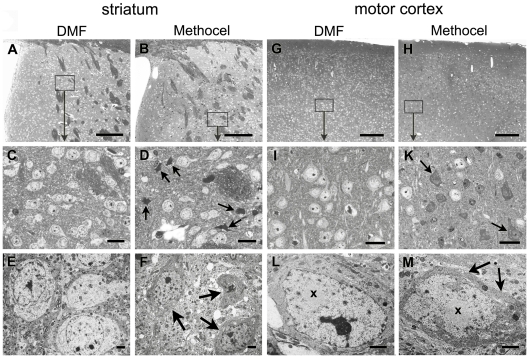
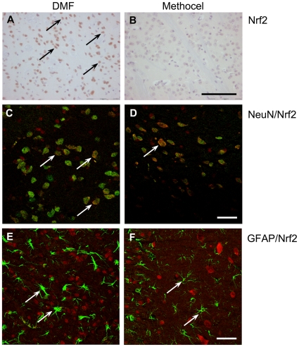
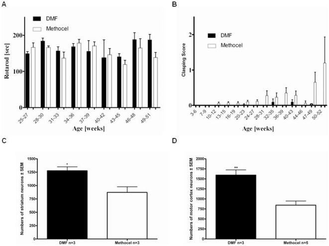
Huntington's disease (HD) is an autosomal dominantly inherited progressive neurodegenerative disease. The exact sequel of events finally resulting in neurodegeneration is only partially understood and there is no established protective treatment so far. Some lines of evidence speak for the contribution of oxidative stress to neuronal tissue damage. The fumaric acid ester dimethylfumarate (DMF) is a new disease modifying therapy currently in phase III studies for relapsing-remitting multiple sclerosis. DMF potentially exerts neuroprotective effects via induction of the transcription factor "nuclear factor E2-related factor 2" (Nrf2) and detoxification pathways. Thus, we investigated here the therapeutic efficacy of DMF in R6/2 and YAC128 HD transgenic mice which mimic many aspects of HD and are characterized by an enhanced generation of free radicals in neurons. Treatment with DMF significantly prevented weight loss in R6/2 mice between postnatal days 80-90. At the same time, DMF treatment led to an attenuated motor impairment as measured by the clasping score. Average survival in the DMF group was 100.5 days vs. 94.0 days in the placebo group. In the histological analysis on day 80, DMF treatment resulted in a significant preservation of morphologically intact neurons in the striatum as well as in the motor cortex. DMF treatment resulted in an increased Nrf2 immunoreactivity in neuronal subpopulations, but not in astrocytes. These beneficial effects were corroborated in YAC128 mice which, after one year of DMF treatment, also displayed reduced dyskinesia as well as a preservation of neurons. In conclusion, DMF may exert beneficial effects in mouse models of HD. Given its excellent side effect profile, further studies with DMF as new therapeutic approach in HD and other neurodegenerative diseases are warranted.


Conflict of interest statement

Competing Interests: The authors have declared that no competing interests exist.

Figures

Figure 1
Figure 1. R6/2 mice display higher levels of oxidative stress.
(A, B) Representative confocal laser scanning image from the striatum of a 3 months old R6/2 mouse (male, A) after hydroethidine staining in comparison to the striatum of a 4 months old C57BL/6 mouse (B). (C, D) Hydroethidine staining of the motor cortex. The same representative R6/2 mouse (C) and C57BL/6 control mouse (D) as in A,B are shown. The amount of free radicals in neuronal cells is indicated in red. Note the increased immunofluorescence in the R6/2 mouse indicating a higher amount of oxidative stress in both anatomical regions. Bar = 20 µm.
Figure 2
Figure 2. DMF improves survival and prevents weight loss in R6/2 mice.
(A) Comparison of body weight changes in DMF treated male R6/2 mice (black bars) and controls treated with carrier solution alone (white bars). Treatment with DMF prevented weight loss in R6/2 mice on postnatal days 85 (p = 0.05) and 90 (p = 0.04). Data are shown as change in body weight (g) on days 80, 85 and 90 to the respective baseline weight at the start of treatment. Numbers of mice per day of analysis are indicated in the graph. (B) Kaplan Meier survival analysis of male R6/2 mice treated with DMF (n = 14, black curve) or methocel (n = 19, grey curve). DMF treatment leads to prolonged survival of R6/2 mice. Confidence interval for mean survival: DMF 97.7–104.6 vs. methocel 91.22–99.6 days.
Figure 3
Figure 3. DMF preserves motor functions in R6/2 mice.
(A) Rotarod analysis. A cohort of R6/2 mice treated with DMF (n = 46) or methocel (n = 42) is shown. Although DMF treatment (black bars) leads to a trend towards longer times on the accelerating rod, there is no significant difference as compared to methocel treated controls (white bars). (B) Representative images of a R6/2 mouse treated with DMF and a mouse treated with methocel at the age of 12 weeks. Note the clasping of the front- and hindlimbs in the control mouse. (C) Clasping Score. A cohort of R6/2 mice treated with DMF (n = 46, black bars) or methocel (n = 42, white bars) is shown. At the age of 12 weeks, there is a significant difference between both groups and DMF treated mice display a reduction of limb dyskinesia.
Figure 4
Figure 4. DMF leads to preservation of morphologically intact neurons.
Representative images of the striatum (A,B and E,F) or motor cortex (C,D and G,H) from DMF (A,C,E,G) or methocel treated mice (B,D,F,H) on day 80 are shown. Bar = 100 µm. Arrows indicate intact, arrowheads indicate degenerating neurons. (A–D) NeuN staining of the striatum (A,B) and the motor cortex (C,D) in R6/2 mice (day 80). Note the higher number of NeuN immunoreactive neurons after DMF treatment (marked by arrows, arrowheads indicate degenerating striatal neurons with loss of NeuN immunoreactivity). (E–H) Cresyl violet staining of the striatum (E,F) and motorcortex (G,H) in R6/2 mice. Note the higher number of intact neurons after DMF treatment (marked by arrows) and the increased number of shrunken, dystrophic neurons after methocel treatment (marked by arrowheads). False color images depicting cresyl violet staining in red are shown.
Figure 5
Figure 5. DMF treatment prevents dark cell degeneration in R6/2 mice.
(A,B) Overview images of semithin sections show the striatum of DMF (A) and methocel treated (B) R6/2 mice at 80 days of age. Representative images are shown. (C,D) At higher enlargement (for localization see rectangles), the light striatal neurons appear intact in the DMF treated mouse (C) whereas dark cells (arrows) are dispersed among intact neurons in the methocel treated mouse (D). (E,F) Electron microscopy confirms neuronal integrity in DMF treated mice (E) and the condensed and shrunken appearance of severely affected striatal neurons in methocel treated mice (F). (G,H) Semithin overviews display regular cortical morphology of the DMF treated mouse (G), but an irregular appearance in the cortex of representative methocel treated mouse (H). (I,K) At higher enlargements (rectangles) of corresponding layers the DMF treated mouse exhibits intact neurons (I) whereas the methocel treated mouse show multiple dark cells of varying size (arrows) dispersed between single intact neurons (K). (L,M) Electron microscopy confirms the dark cell degeneration with shrunken dark cytoplasm (arrows) and a ruffled nuclear envelope of a respective neuron (M) in contrast to the intact cortical neuron of a DMF treated mouse (L). Both the intact and the dark neuron exhibit a prominent intranuclear round inclusion (x). Bars in A,B,G,H = 100 µm; bars in C,D,I,K = 20 µm; bars in E,F,L,M = 1 µm.
Figure 6
Figure 6. Increased Nrf2 immunoreactivity after DMF treatment.
Representative images of the striatum from DMF (A, C, E) or methocel treated mice (B, D, F) on day 80 are shown. (A,B) In contrast to methocel treated R6/2 mice, there is an increased number of Nrf2 positive cells after DMF treatment (Nrf2 immunopositive cells are marked by arrows). Bar = 100 µm. (C,D) Confocal laser scanning microscopy images after NeuN/Nrf2 double staining. In contrast to vehicle treated mice (D), a higher number of NeuN/Nrf2 double positive cells are observed in the striatum in DMF treated R6/2 mice (C). Arrows mark NeuN/Nrf2 double labelled cells. Bar = 20 µm. (E,F) Confocal laser scanning microscopy images after GFAP/Nrf2 double labelling. In the striatum, significant numbers of Nrf2 immunopositive astrocytes were neither identified after DMF treatment (E), nor in control mice (F). Arrows indicate Nrf2 negative astrocytes. Bar = 20 µm; images represent a Z-stack of 10 µm.
Figure 7
Figure 7. DMF preserves motor functions in YAC128 mice.
(A) Rotarod analysis. A cohort of YAC128 mice treated with DMF (n = 23, black bars) or methocel (n = 22, white bars) is shown. Although DMF treatment leads to a trend towards longer times on the accelerating rod, there is no significant difference between both groups. (B) Clasping Score. A cohort of YAC128 mice treated with DMF (n = 23) or methocel (n = 22) is shown. In comparison to control mice (white bars), there is a trend towards reduced clasping behaviour in the DMF treated group (black bars) from the age of 47 weeks. (C) Blinded quantification of neuronal densities in the striatum after creysl violet staining on day 365 (n = 3 per group). There is a significant preservation of neuronal numbers after DMF treatment (black bar) as compared to methocel treatment (white bar, p = 0.03). (D) Blinded quantification of neuronal densities in the motor cortex after creysl violet staining on day 365 (n = 3/5 per group). There is a significant preservation of neuronal numbers after DMF treatment (black bar) as compared to methocel treatment (white bar, p = 0.004).

References

    1. Vonsattel JP, DiFiglia M. Huntington disease. J Neuropathol Exp Neurol. 1998;57:369–384. - PubMed
    1. Kumar P, Kalonia H, Kuman A. Huntington's disease: pathogenesis to animal models. Pharmacol Rep. 2010;62:1–14. - PubMed
    1. Mrowietz U, Christophers E, Altmeyer P. Treatment of severe psoriasis with fumaric acid esters: scientific background and guidelines for therapeutic use. The German Fumaric Ester Consensus Conference. Br J Dermatol. 1999;141:424–429. - PubMed
    1. Schweckendiek W. Treatment of psoriasis vulgaris. Med Monatsschr. 1959;13:103–104. - PubMed
    1. Linker RA, Lee DH, Stangel M, Gold R. Fumarates for the treatment of multiple sclerosis, potential mechanisms of actions and clinical studies. Expert Rev Neurother. 2008;8:1683–1690. - PubMed