. 2011 Jan 31;6(1):e15937.
doi: 10.1371/journal.pone.0015937.

PDE 7 inhibitors: new potential drugs for the therapy of spinal cord injury

Affiliations

PDE 7 inhibitors: new potential drugs for the therapy of spinal cord injury

Irene Paterniti et al. PLoS One. .

Abstract

Background: Primary traumatic mechanical injury to the spinal cord (SCI) causes the death of a number of neurons that to date can neither be recovered nor regenerated. During the last years our group has been involved in the design, synthesis and evaluation of PDE7 inhibitors as new innovative drugs for several neurological disorders. Our working hypothesis is based on two different facts. Firstly, neuroinflammation is modulated by cAMP levels, thus the key role for phosphodiesterases (PDEs), which hydrolyze cAMP, is undoubtedly demonstrated. On the other hand, PDE7 is expressed simultaneously on leukocytes and on the brain, highlighting the potential crucial role of PDE7 as drug target for neuroinflammation.

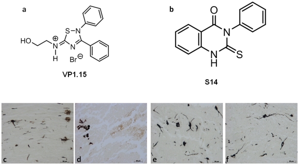
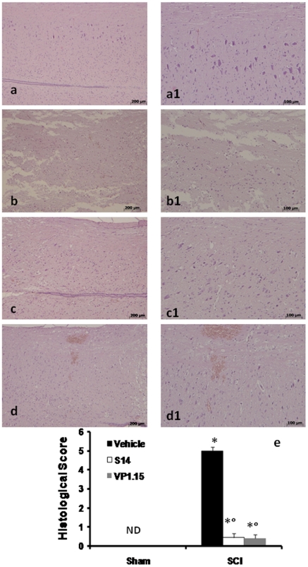
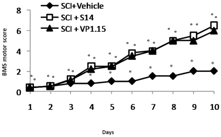
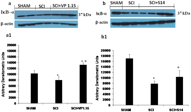
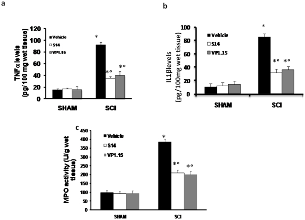
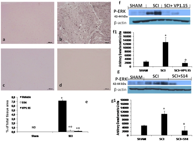
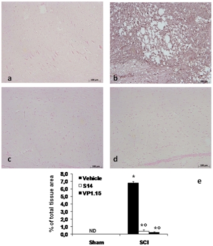
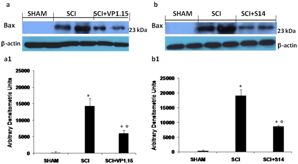
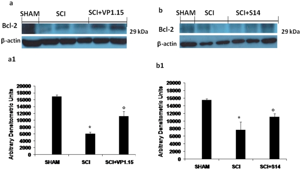
Methodology/principal findings: Here we present two chemically diverse families of PDE7 inhibitors, designed using computational techniques such as virtual screening and neuronal networks. We report their biological profile and their efficacy in an experimental SCI model induced by the application of vascular clips (force of 24 g) to the dura via a four-level T5-T8 laminectomy. We have selected two candidates, namely S14 and VP1.15, as PDE7 inhibitors. These compounds increase cAMP production both in macrophage and neuronal cell lines. Regarding drug-like properties, compounds were able to cross the blood brain barrier using parallel artificial membranes (PAMPA) methodology. SCI in mice resulted in severe trauma characterized by edema, neutrophil infiltration, and production of a range of inflammatory mediators, tissue damage, and apoptosis. Treatment of the mice with S14 and VP1.15, two PDE7 inhibitors, significantly reduced the degree of spinal cord inflammation, tissue injury (histological score), and TNF-α, IL-6, COX-2 and iNOS expression.

Conclusions/significance: All these data together led us to propose PDE7 inhibitors, and specifically S14 and VP1.15, as potential drug candidates to be further studied for the treatment of SCI.


Conflict of interest statement

Competing Interests: The authors have declared that no competing interests exist.

Figures

Figure 1
Figure 1. PDE7 inhibitors and their chemical structures.
a and b represent a chemical structures of VP1,15 and S14respectively. In order in the spinal cord tissue collected from SCI mice, was observed the alteration of morphology of neurons when compared with sham-operated mice (Fig d and c respectively). A protection against alteration of neuron's morphology was observed in mice group treated with VP 1.15 and S14 (e, f respectively).
Figure 2
Figure 2. Effect of PDE7 inhibitors treatment on histological alterations of the spinal cord tissue 24 h after injury.
A significant damage to the spinal cord, from SCI operated mice at the perilesional area, was assessed by the presence of edema as well as alteration of the white matter 24 h after injury (b and b1, see histological score e). Notably, a significant protection from SCI damage was observed in the tissue collected from VP1.15 and S14 treated mice (c, c1 and d, d1 respectively, see histological score e). The histological score (e) was made by an independent observer. wm: White matter; gm: gray matter. This figure is representative of at least 3 experiments performed on different experimental days. Values shown are mean ± s.e. mean of 10 mice for each group. *p<0.01 vs. Sham. °p<0.01 vs. SCI+vehicle.
Figure 3
Figure 3. Effect of VP1.15 and S14 treatment on hind limb motor disturbance after spinal cord injury.
The degree of motor disturbance was assessed every day until 10 days after SCI, by using the Basso mouse scale (BMS) open-field score. Treatment with VP1.15 and S14 reduces the motor disturbance after SCI. Values shown are mean ± s.e. mean of 10 mice for each group. *p<0.01 vs. Sham. °p<0.01 vs. SCI+vehicle.
Figure 4
Figure 4. Effects of VP1.15 and S14 treatment on IκB-α degradation.
By Western Blot analysis, a basal level of IκB-α was detected in the spinal cord from sham-operated animals, whereas IκB-α levels were substantially reduced in SCI mice (a, a1 and b,b1). VP 1.15 and S14 treatment prevented the SCI-induced IκB-α degradation (a, a1 and b,b1 respectively). A representative blot of lysates obtained from each group is shown, and densitometry analysis of all animals is reported. The relative expression of the protein bands from three separated experiments was standardized for densitometry analysis *P<0.01 vs. Sham; °P<0.01 vs. SCI.
Figure 5
Figure 5. Effect of VP1.15 and S14 on TNF-α and IL-1β release and on MPO activity.
A substantial increase in TNF-α (a) and IL-1β (b) production was found in spinal cord tissue collected from SCI mice at 24 h. Spinal cord levels of TNF-α and IL-1β were significantly attenuated by the VP 1.15 and S14 treatment (a, b respectively). Moreover, MPO activity in spinal cord of untreated SCI-operated mice was significantly increased at 24 h after the damage in comparison to sham mice (c). Treatment with VP 1.15 and S14 significantly reduced the SCI-induced increase in MPO activity (c). Data are means ±S.E. mean of 10 mice for each group. *p<0.01 vs sham, °p<. 0.01 vs SCI.
Figure 6
Figure 6. Effects of VP1.15 and S14 on TNF-α and IL-1β expression.
In addition, spinal cord sections were processed at 24 h after SCI to determine the immunohistological staining for TNF-α and IL-1β expression. A substantial increase in TNF-α (b) and IL-1β (f) expression was found in inflammatory cells of the spinal cord tissues from SCI mice at 24th hour after SCI. Spinal cord levels of TNF-α and IL-1β expression were significantly attenuated in VP 1.15 (d,h respectively) and S14 (c, g respectively) treated mice in comparison to SCI animals. Densitometry analysis of immunocytochemistry photographs (n = 5 photos from each sample collected from all mice in each experimental group) for TNF-α and IL-1β (i) from spinal cord tissues was assessed. The assay was carried out by using Optilab Graftek software on a Macintosh personal computer (CPU G3-266). Data are expressed as % of total tissue area. This figure is representative of at least 3 experiments performed on different experimental days. *p<0.01 vs. Sham. °p<0.01 vs SCI+vehicle. ND: not detectable
Figure 7
Figure 7. Effect of VP 1.15 and S14 on activated kinases.
At 24 h after injury we assessed, by immunohistological staining, an increase of Cox-2 expression. A substantial increase in Cox-2 expression was found in spinal cord tissue collected from SCI mice group (b),whereas treatment with VP1.15 (c) and S14 (d) reduced markedly a staining for Cox-2. Densitometry analysis of immunocytochemistry photographs (n = 5 photos from each sample collected from all mice in each experimental group) for Cox-2 (e) from spinal cord tissues was assessed. The assay was carried out by using Optilab Graftek software on a Macintosh personal computer (CPU G3-266). Data are expressed as % of total tissue area. This figure is representative of at least 3 experiments performed on different experimental days. *p<0.01 vs. Sham. °p<0.01 vs SCI+vehicle. ND: not detectable. SCI caused an increase of the ERK1/2 phosphorylation in vehicle-treated mice (f, f1 and g, g1). The treatment with VP1.15 (f, f1) and S14 (g, g1) reduced pERK1/2 levels. Densitometric analysis of protein expression represents the mean ± s.e.mean of 10 spinal cord tissues. Data were normalized on the basis of ERK-2 levels. *P<0.01 vs sham; °P<0.05 vs SCI + vehicle.
Figure 8
Figure 8. Effects of PDE7 inhibitors on iNOS expression.
iNOS expression was evaluated by immunohystochemical analysis in the spinal cord section 24 h after SCI. Spinal cord section from sham-operated mice did not stain for iNOS (a), whereas spinal cord section obtained from SCI-operated mice exhibited positive staining for iNOS (b) mainly localized in various inflammatory cells in the grey matter. VP 1.15 and S14 treatment (c, d respectively) reduced the degree for positive staining for iNOS in the spinal cord tissue. Densitometry analysis of immunocytochemistry photographs (n = 5 photos from each sample collected from all mice in each experimental group) for iNOS (e) from spinal cord tissues was assessed. The assay was carried out by using Optilab Graftek software on a Macintosh personal computer (CPU G3-266). Data are expressed as % of total tissue area. ND: not detectable. This figure is representative of at least 3 experiments performed on different experimental days. *p<0.01 vs Sham. °p<0.01 vs SCI+vehicle.
Figure 9
Figure 9. Western blot analysis for Bax.
By Western blot analysis, Bax levels were appreciably increased in the spinal cord from SCI mice (a, a1 and b, b1). On the contrary, VP1.15 (a, a1) and S14 (b, b1) treatment prevented the SCI-induced Bax expression. The relative expression of the protein bands was standardized for densitometric analysis to β-actin levels, and reported in panel a, a1 and b, b1 are expressed as mean ± s.e.m. from n = 5/6 spinal cord for each group. *P<0.01 vs sham, °P<0.01 vs SCI+vehicle.
Figure 10
Figure 10. Western blot analysis for Bcl-2.
Moreover, a basal level of Bcl-2 expression was detected in spinal cord from sham-operated mice (a, a1 and b, b1). Twenty-four hours after SCI, Bcl-2 expression was significantly reduced in spinal cord from SCI mice (a, a1 and b, b1). Treatment with VP 1.15 (a, a1) and S14 (b, b1) significantly reduced the SCI-induced inhibition of Bcl-2 expression. The relative expression of the protein bands was standardized for densitometric analysis to β-actin levels, and reported in panel a, a1 and b, b1 are expressed as mean ± s.e.m. from n = 5/6 spinal cord for each group. *P<0.01vs sham, °P<0.01 vs SCI+vehicle.
Figure 11
Figure 11. PAMPA-BBB assay results.
A linear correlation between experimental and reported permeability of selected commercial drugs using the experimental conditions described in the PAMPA-BBB assay methodology was found (10a). The permeability (Pe 10−6 cm s−1) value obtained in the PAMPA-BBB assay for the 10 commercial drugs and used in the experiment validation, were reported in Figure 10b. The Pe obtained in the same experimental conditions for compounds S14 and VP1.15 predict their penetration in the CNS.

Similar articles

Cited by

References

    1. Maegele M, Gregor S, Steinhausen E, Bouillon B, Heiss MM, et al. The long-distance tertiary air transfer and care of tsunami victims: injury pattern and microbiological and psychological aspects. Crit Care Med. 2005;33:1136–1140. - PubMed
    1. Bartholdi D, Schwab ME. Methylprednisolone inhibits early inflammatory processes but not ischemic cell death after experimental spinal cord lesion in the rat. Brain Res. 1995;672:177–186. - PubMed
    1. Levy ML, Chen JC, Amar AP, Yamada S, Togo K, et al. Virtual endoscopic environments in modern neurosurgical practice. Neurosurg Focus. 1999;6:e11. - PubMed
    1. Carlson SL, Parrish ME, Springer JE, Doty K, Dossett L. Acute inflammatory response in spinal cord following impact injury. Exp Neurol. 1998;151:77–88. - PubMed
    1. de Castro R, Hughes MG, Xu GY, Clifton C, Calingasan NY, et al. Evidence that infiltrating neutrophils do not release reactive oxygen species in the site of spinal cord injury. Exp Neurol. 2004;190:414–424. - PubMed

Publication types

Substances