. 2011 Jan 31;6(1):e16165.
doi: 10.1371/journal.pone.0016165.

Modulation of Mrp1 (ABCc1) and Pgp (ABCb1) by bilirubin at the blood-CSF and blood-brain barriers in the Gunn rat

Affiliations

Modulation of Mrp1 (ABCc1) and Pgp (ABCb1) by bilirubin at the blood-CSF and blood-brain barriers in the Gunn rat

Silvia Gazzin et al. PLoS One. .

Abstract

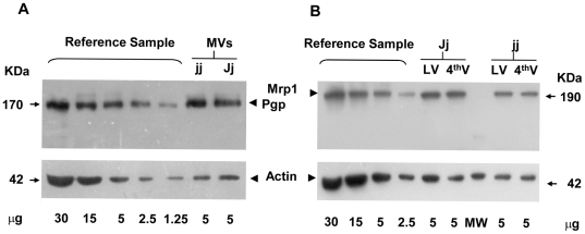
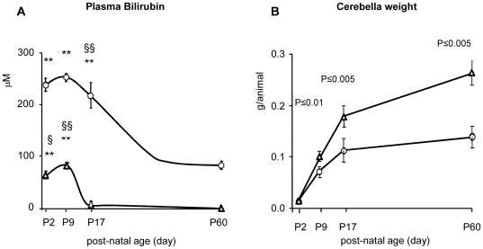
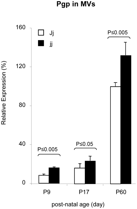
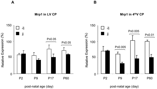
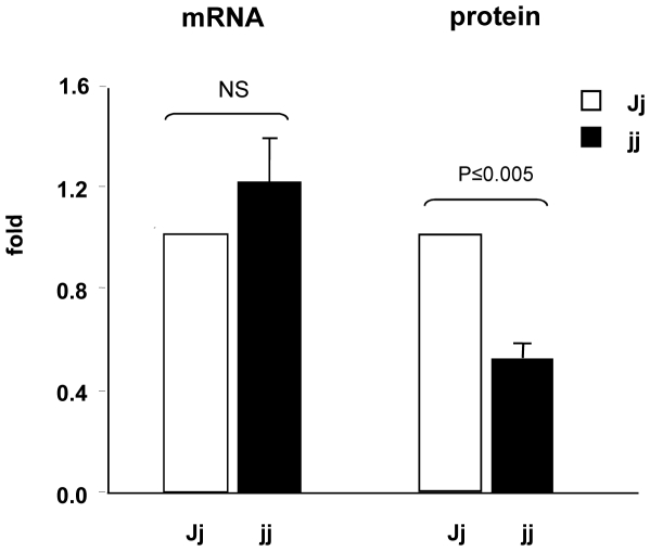
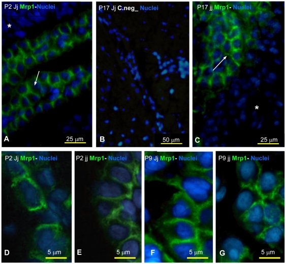
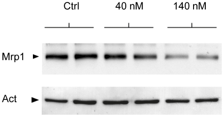
Accumulation of unconjugated bilirubin (UCB) in the brain causes bilirubin encephalopathy. Pgp (ABCb1) and Mrp1 (ABCc1), highly expressed in the blood-brain barrier (BBB) and blood-cerebrospinal fluid barrier (BCSFB) respectively, may modulate the accumulation of UCB in brain. We examined the effect of prolonged exposure to elevated concentrations of UCB on expression of the two transporters in homozygous, jaundiced (jj) Gunn rats compared to heterozygous, not jaundiced (Jj) littermates at different developmental stages (2, 9, 17 and 60 days after birth). BBB Pgp protein expression was low in both jj and Jj pups at 9 days (about 16-27% of adult values), despite the up-regulation in jj animals (2 and 1.3 fold higher than age matched Jj animals at P9 and P17-P60, respectively); Mrp1 protein expression was barely detectable. Conversely, at the BCSFB Mrp1 protein expression was rather high (60-70% of the adult values) in both jj and Jj at P2, but was markedly (50%) down-regulated in jj pups starting at P9, particularly in the 4(th) ventricle choroid plexuses: Pgp was almost undetectable. The Mrp1 protein down regulation was accompanied by a modest up-regulation of mRNA, suggesting a translational rather than a transcriptional inhibition. In vitro exposure of choroid plexus epithelial cells obtained from normal rats to UCB, also resulted in a down-regulation of Mrp1 protein. These data suggest that down-regulation of Mrp1 protein at the BSCFB, resulting from a direct effect of UCB on epithelial cells, may impact the Mrp1-mediated neuroprotective functions of the blood-cerebrospinal fluid barrier and actually potentiate UCB neurotoxicity.


Conflict of interest statement

Competing Interests: The authors have declared that no competing interests exist.

Figures

Figure 1
Figure 1. Representative Western blot for Pgp, Mrp1 and Actin on Gunn preparations.
Pgp expression was analyzed on microvessels (MVs) preparations (A, upper panel). Mrp1 was quantified in lateral and 4th ventricle (LV and 4thV) choroid plexuses (B, upper panel). Actin staining (both A and B, lower panel) was used to normalize the amount of samples loaded. For the quantification procedure, see the material and methods section.
Figure 2
Figure 2. Plasma bilirubin and cerebellar weight in jj and Jj Gunn rats.
The values are expressed as mean ± SD of 12–20 animals for each age and genotype. Round filled dots: jj animals; empty triangles: Jj controls. Panel A: total plasma bilirubin (Tbil) amount in developing hyperbilirubinemic (jj) and control (Jj) Gunn rats. Tbil concentration differences between jj and age-matched Jj animals were all statistically significant (P≤0.005). Within each phenotype: **: p<0.005 vs. P60; § and §§ : p<0.05 and 0.005 respectively vs. next age group. Panel B shows the cerebellar growth expressed as g/animal. The level of significance is indicated by comparison with age-matched not jaundiced Jj animals.
Figure 3
Figure 3. Blood brain barrier localization in rat brain, and microvessels isolated from rats.
Cartoon representation of the lateral, 3rd, 4th ventricle choroid plexuses (LV CP, 3rd V CP and 4thV CP, respectively) and microvessels (MVs) localization in rat brain (A). Representative microvessels freshly isolated from rat brains. Freshly isolate MVs in 0.1% albumin in Krebbs-Ringer buffer were placed on a microscope slide and directly observed by phase contrast microscopy: In B scale bar 100 µm; in C, D, E, F and G scale bar 25 µm. P: post-natal age in days. jj: hyperbilirubinemic rats; Jj: controls.
Figure 4
Figure 4. Pgp relative expression in isolated Jj and jj Gunn rat microvessels.
The Pgp amount in each sample is expressed as % of the Pgp amount in the Reference Sample (Pooled P60 Jj MVs preparations). White bars: heterozygous (Jj) rats, black bars: hyperbilirubinemic homozygous (jj) animals. The values are expressed as mean ± SD. Statistical significance is indicated.
Figure 5
Figure 5. Mrp1 relative expression in the lateral and 4th ventricle choroid plexuses dissected from Jj and jj Gunn rats.
The Mrp1 protein amount present in each sample is expressed as % of the Mrp1 amount in the Reference Sample (pooled P60 Jj 4thV CP preparations). White bars: heterozygous (Jj) rats, black bars: hyperbilirubinemic homozygous (jj) animals. 4thV CP: Forth Ventricle Choroid Plexuses; LV CP: Lateral Ventricles Choroid Plexuses. Statistical significance is indicated.
Figure 6
Figure 6. Comparison of Mrp1 mRNA and protein relative expression in choroid plexuses.
White bars: heterozygous (Jj) P9 rats, black bars: hyperbilirubinemic homozygous (jj) P9 animals. The values are expressed as mean ± SD. Statistical significance is reported. NS: not significant difference.
Figure 7
Figure 7. Immunofluorescence for Mrp1 on Gunn rat brain slices during post-natal development.
A, B, D, F: heterozygous (Jj) animals. C, E, G: hyperbilirubinemic homozygous (jj) animals. A negative control obtained omitting the first antibody is shown in B. A and C, show the specific expression of Mrp1 at the blood-cerebrospinal fluid barrier of Jj and jj Gunn rats. Green: Mrp1;Blue: Nuclei; → : basolateral side; * : parenchyma. In D, E (P2 Jj vs jj) and F,G (P9 Jj vs jj) magnifications showing Mrp1 localization at the basolateral side of choroidai plexus cells.
Figure 8
Figure 8. Western blot analysis of Mrp1 protein in the choroidal epithelium following bilirubin treatment.
Duplicate monolayers of choroidal epithelial cells were included for each experimental condition. Following daily basolateral treatment with 40 or 140 nM unbound bilirubin for 6 consecutive days, filters were treated in exactly the same conditions. The homogeneity in protein loading is reflected by the actin band revealed on the lower part of the membrane.

Similar articles

Cited by

References

    1. Gourley GR. Bilirubin metabolism and kernicterus. Adv Pediatr. 1997;44:173–229. - PubMed
    1. Ostrow JD, Pascolo L, Shapiro SM, Tiribelli C. New concepts in bilirubin encephalopathy. Eur J Clin Invest. 2003;33:988–997. - PubMed
    1. Ostrow JD. Bile pigments and jaundice. Marcel Dekker, Inc; 1987.
    1. Kaplan M, Muraca M, Hammerman C, Rubaltelli F, Vilei MT, et al. Imbalance between production and conjugation of bilirubin: a fundamental concept in the mechanism of neonatal jaundice. 2002. Available: ttp://pediatrics.org/cgi/content/full/110/4/e47. - PubMed
    1. Shapiro SM, Bhutani VK, Johnson L. Hyperbilirubinemia and kernicterus. Clin Perinatol. 2006;33:387–410. - PubMed

Publication types

MeSH terms

Substances