. 2011 Jan 28;6(1):e16458.
doi: 10.1371/journal.pone.0016458.

Primary human mDC1, mDC2, and pDC dendritic cells are differentially infected and activated by respiratory syncytial virus

Affiliations

Primary human mDC1, mDC2, and pDC dendritic cells are differentially infected and activated by respiratory syncytial virus

Teresa R Johnson et al. PLoS One. .

Abstract

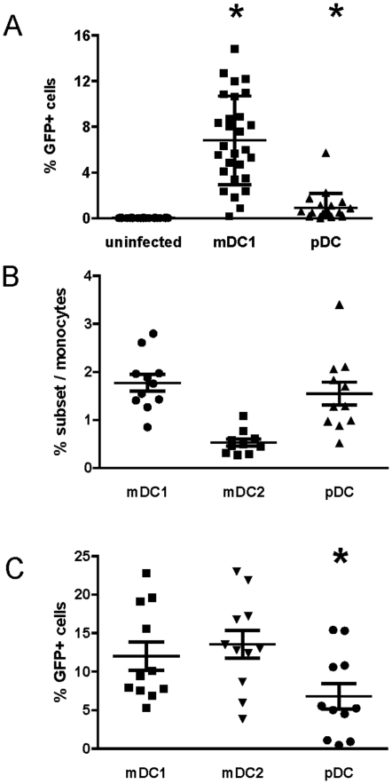
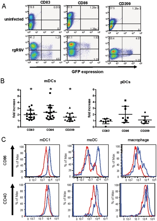
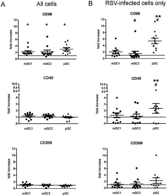
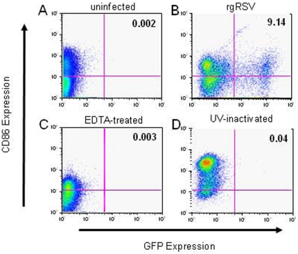
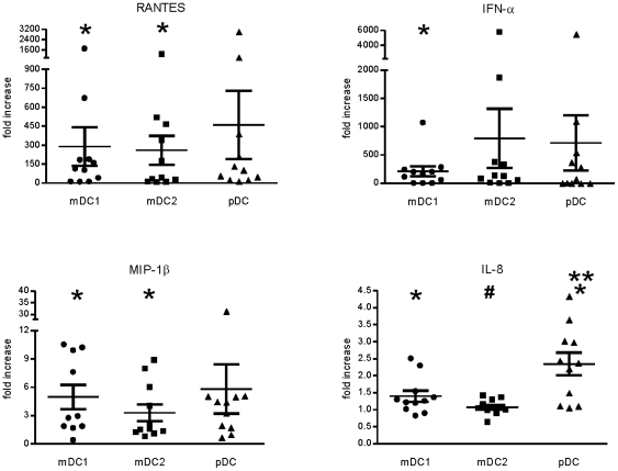
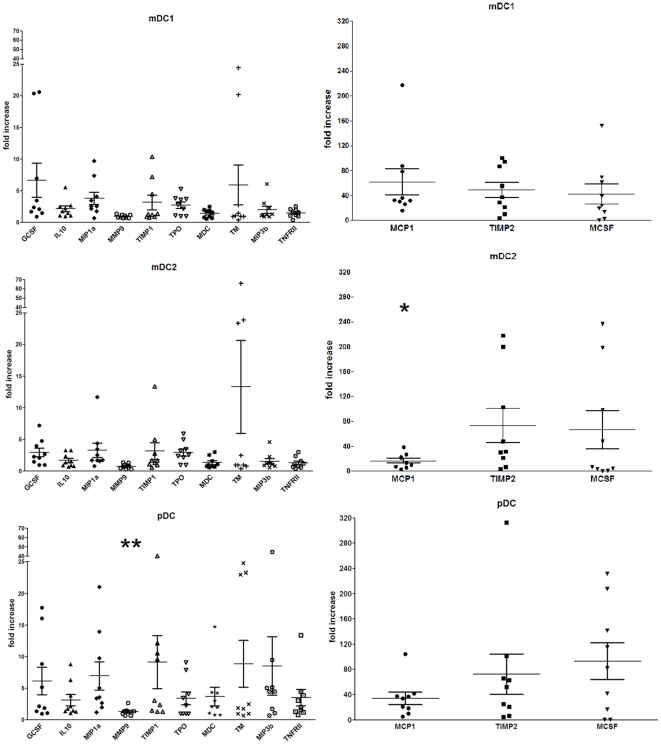
Respiratory syncytial virus (RSV) causes recurrent infections throughout life. Vaccine development may depend upon understanding the molecular basis for induction of ineffective immunity. Because dendritic cells (DCs) are critically involved in early responses to infection, their interaction with RSV may determine the immunological outcome of RSV infection. Therefore, we investigated the ability of RSV to infect and activate primary mDCs and pDCs using recombinant RSV expressing green fluorescent protein (GFP). At a multiplicity of infection of 5, initial studies demonstrated ∼6.8% of mDC1 and ∼0.9% pDCs were infected. We extended these studies to include CD1c(-)CD141(+) mDC2, finding mDC2 infected at similar frequencies as mDC1. Both infected and uninfected cells upregulated phenotypic markers of maturation. Divalent cations were required for infection and maturation, but maturation did not require viral replication. There is evidence that attachment and entry/replication processes exert distinct effects on DC activation. Cell-specific patterns of RSV-induced maturation and cytokine production were detected in mDC1, mDC2, and pDC. We also demonstrate for the first time that RSV induces significant TIMP-2 production in all DC subsets. Defining the influence of RSV on the function of selected DC subsets may improve the likelihood of achieving protective vaccine-induced immunity.


Conflict of interest statement

Competing Interests: The authors have declared that no competing interests exist.

Figures

Figure 1
Figure 1. Frequency and RSV infection rates of primary mDC1, mDC2 and pDC.
Panel A – Primary pDC and mDC1 were sequentially isolated from elutriated human monocytes in the first cohort of donors by magnetic selection and infected with rgRSV (moi = 5) as described in the Materials and Methods. The percentage of infected (GFP+) cells was determined 18–24 hrs post-infection for mock-infected cells (•, n = 27), mDC1 (▪, n = 27), and pDCs (▴, n = 20). Relative to mock-infected cells, significantly more mDC1 and pDCs are GFP+ (p<0.0001 and p = 0.007, respectively). Panel B – In a second cohort of donors, pDC, mDC1, and mDC2 were sequentially isolated from elutriated monocytes. The cell subsets were counted and frequency within the elutriated monocytes calculated. Panel C – Following magnetic selection and overnight resting, mDC1, mDC2, and pDC were exposed to live rgRSV (moi = 5). Twenty-four hours after infection, uninfected and infected cells were fixed and analyzed by flow cytometry. Data are represented as the percentage of GFP+ cells within live DCs. For panels B and C, N = 11 donor samples with 1 donor present for 2 separate visits. *  =  significantly different from mDC1 and mDC2, p<0.05.
Figure 2
Figure 2. RSV infection and maturation of primary mDC1 and pDCs and of moDCs and macrophages.
From elutriated monocytes of a single donor, primary mDC1 and pDC were isolated and moDC and macrophages were generated as detailed in the Materials and Methods. Panel A – Isolated mDC1 (shown above) and pDC (not shown here) were infected with rgRSV (moi = 5). RSV infection (GFP expression) and DC maturation (CD83, CD86, and CD209 expression) were evaluated after overnight incubation with rgRSV. Dot plot results for mDC1 from a single representative donor (of 15) are shown above. Panel B – mDC1 and pDCs were exposed to live RSV, and DC maturation was examined 18–24 hrs later. Expression of each maturation marker in mDC1 and pDCs is shown, normalizing the mean fluorescence intensity (MFI) in RSV-exposed DC cultures to mock-infected DCs for the same donor. N = 16 for mDC1 and 8 for pDCs. Panel C – After overnight exposure of primary mDC1, moDC, and macrophages from a single donor to live rgRSV, DC maturation was examined by flow cytometric analyses of CD86 and CD40 expression. A representative maturation profile is shown with similar maturation patterns observed in 5 independent donors.
Figure 3
Figure 3. Maturation of RSV-exposed mDC1, mDC2, and pDCs.
DC subsets were isolated and infected as detailed in Figure 1. Twenty-four hours after RSV exposure, cells were stained for expression of surface maturation markers and analyzed by flow cytometry. Data are represented as the mean channel fluorescence of RSV-exposed DCs normalized to the mean channel fluorescence of mock-infected cell controls, gating on all live DCs (panel A) or on live GFP+ cells only (panel B). Actual (non-normalized) MFI values for each donor are detailed in Table S3. N = 11 with 1 donor present for 2 separate visits. *  =  significantly different from mock-infected cell of the same type; **  =  significantly different from mDC1 and mDC2; #  =  significantly different comparing total cells and GFP+ cells only. p<0.05.
Figure 4
Figure 4. RSV infection of mDC1 requires divalent cations and viral replication, but maturation does not require replication.
mDC1 were mock infected (panel A), or exposed to live RSV (panel B), RSV treated with 0.05 M EDTA (panel C), or UV-inactivated RSV (panel D). Equivalent viral particles (moi = 5 of live virus) were used for each RSV exposure. Twenty-four hours after virus exposure the cells were stained for maturation markers (CD83, CD86, CD209) and analyzed by flow cytometry. GFP and CD86 expression is shown in a representative donor (of 8) in which both EDTA-treated and UV-inactivated RSV were used. Data for all donors are summarized in Table 1.
Figure 5
Figure 5. Cytokine and chemokine production as measured by standard sandwich ELISA.
DC subsets were isolated and exposed to RSV as described in Figure 1. Twenty-four hours after RSV infection, the culture supernatants were removed and frozen. Subsequently, cytokine and chemokine levels were measured by sandwich ELISA according to kit protocols. Data represent production levels of RSV-exposed DCs normalized to uninfected DCs of the same cell type. Actual (non-normalized) cytokine concentrations for each donor are detailed in Table S3. N = 11 with 1 donor present for 2 separate visits. *  =  significantly different from mock-infected cells of the same type; **  =  significantly different from mDC1 and mDC2; #  =  significantly different from mDC1 only. p<0.05.
Figure 6
Figure 6. RSV-induced of cytokine, chemokine, and growth factor production measured by multiplex analyses.
Cytokine and chemokine levels were measured by multiplex assay. Data represent production levels of RSV-exposed DCs normalized to uninfected DCs of the same cell type. Actual (non-normalized) cytokine concentrations for each donor are detailed in Table S3. N = 9 unique donors. *  =  significantly different from mDC1 only; **  =  significantly different from mDC1 and mDC2, p<0.05.

Similar articles

Cited by

References

    1. Collins PL, Graham BS. Viral and host factors in human respiratory syncytial virus pathogenesis. J Virol. 2008;82:2040–2055. - PMC - PubMed
    1. Martinez FD. Respiratory syncytial virus bronchiolitis and the pathogenesis of childhood asthma. Pediatr Infect Dis J. 2003;22:S76–S82. - PubMed
    1. Schauer U, Hoffjan S, Bittscheidt J, Köchling A, Hemmis S, et al. RSV bronchiolitis and risk of wheeze and allergic sensitisation in the first year of life. Eur Respir J. 2002;20:1277–1283. - PubMed
    1. Sigurs N, Bjarnason R, Sigurbergsson F, Kjellman B. Respiratory syncytial virus bronchiolitis in infancy is an important risk factor for asthma and allergy at age 7. Am J Resp Crit Care Med. 2000;161:1501–1507. - PubMed
    1. Glezen WP, Taber LH, Frank AL, Kasel JA. Risk of primary infection and reinfection with respiratory syncytial virus. Am J Dis Child. 1986;140:543–546. - PubMed

Publication types

MeSH terms

Substances