. 2011 Jan 27;6(1):e16544.
doi: 10.1371/journal.pone.0016544.

Allelic diversity of the Plasmodium falciparum erythrocyte membrane protein 1 entails variant-specific red cell surface epitopes

Affiliations

Allelic diversity of the Plasmodium falciparum erythrocyte membrane protein 1 entails variant-specific red cell surface epitopes

Inès Vigan-Womas et al. PLoS One. .

Abstract

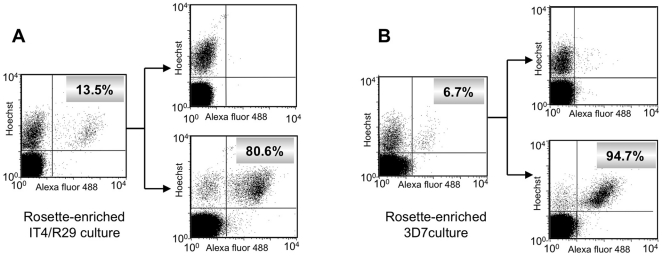
The clonally variant Plasmodium falciparum PfEMP1 adhesin is a virulence factor and a prime target of humoral immunity. It is encoded by a repertoire of functionally differentiated var genes, which display architectural diversity and allelic polymorphism. Their serological relationship is key to understanding the evolutionary constraints on this gene family and rational vaccine design. Here, we investigated the Palo Alto/VarO and IT4/R29 and 3D7/PF13_003 parasites lines. VarO and R29 form rosettes with uninfected erythrocytes, a phenotype associated with severe malaria. They express an allelic Cys2/group A NTS-DBL1α(1) PfEMP1 domain implicated in rosetting, whose 3D7 ortholog is encoded by PF13_0003. Using these three recombinant NTS-DBL1α(1) domains, we elicited antibodies in mice that were used to develop monovariant cultures by panning selection. The 3D7/PF13_0003 parasites formed rosettes, revealing a correlation between sequence identity and virulence phenotype. The antibodies cross-reacted with the allelic domains in ELISA but only minimally with the Cys4/group B/C PFL1955w NTS-DBL1α. By contrast, they were variant-specific in surface seroreactivity of the monovariant-infected red cells by FACS analysis and in rosette-disruption assays. Thus, while ELISA can differentiate serogroups, surface reactivity assays define the more restrictive serotypes. Irrespective of cumulated exposure to infection, antibodies acquired by humans living in a malaria-endemic area also displayed a variant-specific surface reactivity. Although seroprevalence exceeded 90% for each rosetting line, the kinetics of acquisition of surface-reactive antibodies differed in the younger age groups. These data indicate that humans acquire an antibody repertoire to non-overlapping serotypes within a serogroup, consistent with an antibody-driven diversification pressure at the population level. In addition, the data provide important information for vaccine design, as production of a vaccine targeting rosetting PfEMP1 adhesins will require engineering to induce variant-transcending responses or combining multiple serotypes to elicit a broad spectrum of immunity.


Conflict of interest statement

Competing Interests: The authors have declared that no competing interests exist.

Figures

Figure 1
Figure 1. Domain organisation of the PfEMP1 proteins and sequence comparison of the NTS-DBL1α domains.
(A) Schematic domain organisation of the Palo Alto/varO, IT4/R29, 3D7/PF13_0003 and 3D7/PFL1955w PfEMP1 proteins. Regions corresponding to the recombinant NTS-DBL1α domains are indicated by bars, with the N- and C-terminal residue numbers giving sequence limits. Domains in dark grey have >50% sequence identity, shaded DBLβ domains have ∼38% identity; DBL1α-PFL1955w is shown in light grey (29–33% identity). (B) The NTS-DBL1α sequences aligned as described in the Materials and Methods. Cysteine residues are shown in white on a black background and residues with 50% or more sequence identity are shown in white on a grey background.
Figure 2
Figure 2. ELISA titration curves of mouse sera against the NTS-DBL1α recombinant domains.
Serial two-fold dilutions of mouse anti-VarO (A), anti-R29 (B), anti-PF13 (C) polyclonal sera (initial dilution 1/200) tested by ELISA against the recombinant domains. The plates were coated with the VarO (squares), R29 (triangles), PF13 (open circles) and PFL1955w (diamonds) domains; (x) indicates pre-immune sera.
Figure 3
Figure 3. Isolation of monovariant cultures of IT4/R29 and 3D7/PF13_0003.
Rosette-enriched IT4/R29 (A) and 3D7/PF13 (B) cultures were incubated with polyclonal sera raised against recombinant domains and were isolated by cell sorting (see Materials and Methods). The left-hand panels show surface positivity rates of the initial rosette-enriched cultures and the right-hand panels show the surface reactivity of the negative (upper) and positive (lower) sorted populations. The x and y axes show Log10 fluorescence.
Figure 4
Figure 4. Variant-specific iRBC surface serotypes of Palo Alto/VarO, IT4/R29 and 3D7/PF13.
(A) Surface immunofluorescence: monovariant lines were incubated with mouse sera raised to the recombinant domain and analysed by FACS. Rows and columns show parasite lines and sera, respectively, as indicated. The x and y axes show Log10 fluorescence. (B) Variant-specific surface reactivity of the mAbs raised to the recombinant domains with the monovariant lines analysed by FACS. In each panel, the shaded area, the thick line and the thin line indicate reactivity with Palo Alto/VarO, IT4/R29 and 3D7/PF13, respectively. (C) Variant-specific rosette disruption. Monovariant cultures were incubated with mouse serum as indicated and the fraction of mature stages forming rosettes was monitored. White, light grey and dark grey bars indicate Palo Alto/VarO, IT4/R29 and 3D7/PF13, respectively.
Figure 5
Figure 5. Seroprevalence for NTS-DBL1α domains and Palo Alto/VarO, IT4/R29 and 3D7/PF13 iRBC in Dielmo (Senegal).
Seroprevalence to recombinant domains (A) and the iRBC surface (B) in Dielmo, Senegal, measured by ELISA and FACS, respectively. The boundaries of the boxes indicate the 25th and 75th percentiles, and the line in each box indicates the median. The whiskers indicate the 10th and 90th percentiles. The outlying dots show values exceeding the 10th and 90th percentiles. The number of persons by age group was 9, 23, 19, 14, 12 and 158 in the 0.5->2, 2-<5, 5-<7, 7-<9, 9-<11 and >11 years, respectively. Symbols used: open, VarO; light grey, R29; medium grey, PF13; dark grey, PFL1955w (in A only).
Figure 6
Figure 6. Mixed agglutination assay with Palo Alto/VarO, IT4/R29 and 3D7/PF13 monovariant lines.
After rosette disruption with dextran sulphate, the RBC membrane was labelled with either PKH26 or PKH67, mixed in homologous or heterologous pairwise association, and incubated with human serum in the presence of dextran sulphate. Agglutination was examined using a fluorescence microscope. A representative example with an individual serum is shown. (A), (B) and (C) show mixed agglutinates of homologous PKH26- and PKH67-labeled Palo Alto/VarO, IT4/R29 and 3D7/PF13 associations, respectively, while (D) shows a typical absence of mixed agglutinates using heterologous association (shown is a PKH26-labeled Palo Alto/VarO and PKH67-labeled IT4/R29 association).
Figure 7
Figure 7. Residual VarO-iRBC surface reactivity after serum depletion of human sera using the recombinant VarO domain.
FACS analysis of serum from adults (A and C) and from a 6 y old child (B) living in Dielmo, Senegal before and after antibody depletion. The grey area indicates fluorescence intensity distribution of non-immune French blood donors. The thick, dotted and thin lines indicate IFA distribution of non-depleted, control TALON-absorbed, recombinant VarO-depleted serum, respectively. The x axis shows Log10 fluorescence.
Figure 8
Figure 8. Sequence polymorphism of the three alleles mapped upon the NTS-DBL1α1 varO crystal structure.
Three mutually orthogonal views of the surface of the NTS-DBL1α1-varO crystal structure with sequence polymorphism between the three alleles, VarO, R29 and PF13, colour-coded from blue (variable) to red (conserved - see the nine-point colour scale, insert). (A) and (C) are viewed with the C-terminus of the domain at the bottom and are rotated with respect to each other by 180° about a vertical axis. (B) is a perpendicular view, seen from above (A) and (C).

Similar articles

Cited by

References

    1. Contamin H, Fandeur T, Rogier C, Bonnefoy S, Konate L, et al. Different genetic characteristics of Plasmodium falciparum isolates collected during successive clinical malaria episodes in Senegalese children. Am J Trop Med Hyg. 1996;54:632–643. - PubMed
    1. Marsh K, Howard RJ. Antigens induced on erythrocytes by P. falciparum: expression of diverse and conserved determinants. Science. 1986;231:150–153. - PubMed
    1. Bull PC, Lowe BS, Kortok M, Molyneux CS, Newbold CI, et al. Parasite antigens on the infected red cell surface are targets for naturally acquired immunity to malaria. Nat Med. 1998;4:358–360. - PMC - PubMed
    1. Smith JD, Chitnis CE, Craig AG, Roberts DJ, Hudson-Taylor DE, et al. Switches in expression of Plasmodium falciparum var genes correlate with changes in antigenic and cytoadherent phenotypes of infected erythrocytes. Cell. 1995;82:101–110. - PMC - PubMed
    1. Kraemer SM, Smith JD. A family affair: var genes, PfEMP1 binding, and malaria disease. Curr Opin Microbiol. 2006;9:374–380. - PubMed

Publication types