. 2011 Jan 27;7(1):e1001262.
doi: 10.1371/journal.ppat.1001262.

A cardinal role for cathepsin d in co-ordinating the host-mediated apoptosis of macrophages and killing of pneumococci

Affiliations

A cardinal role for cathepsin d in co-ordinating the host-mediated apoptosis of macrophages and killing of pneumococci

Martin A Bewley et al. PLoS Pathog. .

Abstract

The bactericidal function of macrophages against pneumococci is enhanced by their apoptotic demise, which is controlled by the anti-apoptotic protein Mcl-1. Here, we show that lysosomal membrane permeabilization (LMP) and cytosolic translocation of activated cathepsin D occur prior to activation of a mitochondrial pathway of macrophage apoptosis. Pharmacological inhibition or knockout of cathepsin D during pneumococcal infection blocked macrophage apoptosis. As a result of cathepsin D activation, Mcl-1 interacted with its ubiquitin ligase Mule and expression declined. Inhibition of cathepsin D had no effect on early bacterial killing but inhibited the late phase of apoptosis-associated killing of pneumococci in vitro. Mice bearing a cathepsin D(-/-) hematopoietic system demonstrated reduced macrophage apoptosis in vivo, with decreased clearance of pneumococci and enhanced recruitment of neutrophils to control pulmonary infection. These findings establish an unexpected role for a cathepsin D-mediated lysosomal pathway of apoptosis in pulmonary host defense and underscore the importance of apoptosis-associated microbial killing to macrophage function.


Conflict of interest statement

The authors have declared that no competing interests exist.

Figures

Figure 1
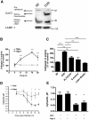
Figure 1. Differentiated THP-1 cells infected with pneumococci exhibit loss of lysosomal acidification and cytosolic translocation of cathepsin D.
Differentiated THP-1 cells were infected with pneumococci (D39) and stained with (A, C) acridine orange (AO) and (B, D) JC-1 at the designated times post infection. (A–B) Representative histograms from one infection and (C–D) graphs summarizing loss of lysosomal acidification (LLA) and inner mitochondrial transmembrane potential (ΔΨm) in three separate experiments are shown, * = p<0.05, ** = p<0.01, one way ANOVA with Dunnett's post-test vs. 0 h. (E) AO staining 16 h post-infection of mock infected (Spn −) or pneumococcal infected (Spn+) differentiated THP-1 cells in the presence (+) or absence (−) of zVADfmk (zVAD) or zFAfmk (zFA), n = 3. Spn+ without zVAD/zFA vs. Spn+ with zVAD, p = ns (not significant) (F) Mock (Spn−) or D39 (Spn+) infected cells were stained with BODIPY FL-Pepstatin A 16 h post-infection and either visualized by microscopy or analyzed by flow cytometry. The filled histogram is Spn−, black histogram is Spn+, grey is unstained (US). The images and flow histograms are representative of three independent experiments. Scale bar equal to 5 µm. Quantified flow results are shown below the histogram, n = 3 (G) A Western blot of cytosolic and membrane fractions from mock (Spn−) or D39 (Spn+) infected differentiated THP-1 cells at 16 h post-infection probed with anti–cathepsin D (CatD) and cathepsin B (CatB). Actin and LAMP-1 were used as loading controls. The blots are representative of three independent experiments. (H) AO staining of differentiated THP-1 cells 16 h after mock-infection (MI) or exposure to D39 or a pneumolysin-deficient strain of D39 (PLYSTOP), n = 4, *** p<0.001, one-way ANOVA with Tukey's post-test. In all cases, pooled data are expressed as mean +/− SEM.
Figure 2
Figure 2. Infection with pneumococci is associated with activation of cathepsin D in differentiated THP-1 cells.
(A) A Western blot of phagolysosomes prepared from differentiated THP-1 cells 14 h after mock infection (MI), or infection with S. pneumoniae strain D39 and isolated using a discontinuous sucrose gradient was probed for cathepsin D. The blot is representative of three independent infections. (B) Cathepsin D activity was measured in whole cell lysates from mock (Spn−) or D39 (Spn+) infected differentiated THP-1 cells at the designated time points. D39 infected cells showed elevated cathepsin D activity compared to mock infected cells from 8 h, n = 4, *** = p<0.001, two-way ANOVA (C) Cathepsin D activity measured in whole cell lysates at 14 h in mock-infected (MI) cells, or differentiated THP-1 cells infected with the designated Spn strains (D39 or the pneumolysin-deficient strain PLYSTOP), Staph. aureus or latex beads, n = 4, ns =  not significant * = p<0.05. ** = p<0.01, *** = p<0.001 one-way ANOVA with Tukey's post-test. (D) Cytosolic pH was measured in mock (Spn−) or D39 (Spn+) infected cells at the designated time points using SNARF-4F carboxylic acid, acetoxymethyl ester, acetate, n = 4, * = p<0.05. ** = p<0.01, two-way ANOVA. (E) Cytosolic pH was measured at 14 h in differentiated THP-1 cells either mock-infected (Spn−) or exposed to D39 pneumococci (Spn+) in the presence (+) or absence (−) of pepstatin A (Pep A), n = 4, * = p<0.05, one-way ANOVA with Dunnett's post-test vs. MI. In all cases, pooled data are expressed as mean +/− SEM.
Figure 3
Figure 3. Cathepsin D activity contributes to apoptosis in the differentiated THP-1 cell line.
(A) Differentiated THP-1 cells were stained with JC-1 16 h after mock-infection (Spn−) or exposure to D39 pneumococci (Spn+) in the presence (+) or absence (−) of pepstatin A or MPC6. Loss of fluorescence indicates loss of ΔΨm, n = 3–5, * = p<0.05, ** = p<0.01, two-way ANOVA. (B) A representative Western blot of the cytosolic fractions of Spn− or Spn+ cells, 16 h after infection, in cultures incubated with (+) or without (−) pepstatin A (PepA). The blot is representative of four independent infections. (C) Spn- or Spn+ differentiated THP-1 cells, incubated in the presence (+) or absence (−) of pepstatin A (PepA), were assayed for caspase activity by fluorimetry 16 h post-infection, n = 5, * = p<0.05. (D) Spn− or Spn+ differentiated THP-1 cells, incubated in the presence (+) or absence (−) of pepstatin A or MPC6, were fixed and analyzed for nuclear fragmentation after 20 h culture, n = 3–4, * = p<0.05, ** = p<0.01, two-way ANOVA. In all cases, pooled data are expressed as mean +/− SEM.
Figure 4
Figure 4. Macrophages deficient in cathepsin D show reduced apoptosis.
(A) BMDM's from (WT) or cathepsin D knock-out (KO) mice were stained with JC-1, 16 h after mock-infection (Spn−) or pneumococcal infection (Spn+) with D39 in the presence (+) or absence (−) of pepstatin A (PepA), n = 7, * = p<0.05, two-way ANOVA. (B) Nuclear fragmentation was detected by DAPI staining, in WT and cathepsin D KO BMDMs, 20 h after mock (Spn−) or D39 pneumococcal (Spn+) infection in the presence (+) or absence (−) of PepA, n = 5 per group. (C) Acridine orange staining of BMDMs 16 h post-infection in the presence (+) or absence (−) of pepstatin A, n = 7 per group, * = p<0.05, two-way ANOVA. In all cases, pooled data are expressed as mean +/− SEM.
Figure 5
Figure 5. Cathepsin D facilitates Mcl-1 downregulation.
(A) A representative Western blot for Mcl-1 from wild-type (WT) and Cathepsin D knock-out (KO) BMDMs 16 h after mock- (Spn−) or D39 pneumococcal-infection (Spn+), in the presence (+) or absence (−) of pepstatin A. The blot is representative of four independent experiments. Densitometry was carried out and fold change of Mcl-1 relative to mock-infection was calculated, n = 4 * = p<0.05, two-way ANOVA. (B) Spn− or Spn+ differentiated THP-1 cells were cultured in the presence (+) or absence (−) of pepstatin A (PepA). At 16 h cells were lysed, ubiquitinated proteins captured, and Western blots carried out probing for total ubiquitinated proteins and for Mcl-1. Densitometry was carried out and the ratios of Mcl-1 relative to ubiquitin were calculated, n = 3, * = p<0.05, one-way ANOVA with Tukey's post-test. (C) Spn− or Spn+ differentiated THP-1 cells were lysed and probed for Hsp70, Mule and actin at the designated times post-infection. The blots are representative of three experiments and the results summarized by densitometry.
Figure 6
Figure 6. Cathepsin D activation favors the interaction between Mcl-1 and Mule.
(A) Mock-infected (Spn−) or D39 exposed (Spn+) differentiated THP-1 cells were immunoprecipitated with an anti-Mcl-1 antibody at the designated time period. As controls a separate sample at each time point was treated with a Mcl-1 peptide. The peptide used was identical to that used to produce the anti-Mcl-1 antibody. This excluded non-specific binding by the anti-Mcl-1 antibody. Precipitated proteins were blotted for Hsp70, Mule and Mcl-1. Densitometry was carried out and the ratios of Mule and Hsp70 to the amount of Mcl-1 precipitated was calculated, n = 3 * = p<0.05 for comparison of 4 h vs. 16 h, 1-way ANOVA with Dunnett's post test. Spn− or Spn+ differentiated THP-1 cells were cultured in the presence (+) or absence (−) of pepstatin A (PepA) for 16 h and lysates were immunoprecipitated with anti-Mcl-1 (B) or anti-Mule (C) antibody before being probed for Mule, Hsp70 and Mcl-1. C represents a control in which mock-infected (MI) cells were immunoprecipitated in the presence of an excess of antigen-specific peptide (Mcl-1) or a non-specific antibody for the immunoprecipitation of Mule. Densitometry was carried out and the ratios of the immunoblotted proteins to the immunoprecipitated Mcl-1 or Mule were calculated, n = 3 *** = p<0.001 for comparison of Spn+ with or without pepstatin A, one-way ANOVA with Tukey's post-test.
Figure 7
Figure 7. Cathepsin D in alveolar macrophages contributes to bacterial killing in vitro and in vivo.
(A) BMDMs from (WT) or cathepsin D knock-out (KO) mice were exposed to D39 pneumococci for the indicated time periods. Cells were lysed and intracellular bacteria plated out at the designated times, n = 5 per group, * = p<0.05, two-way ANOVA. (B) A representative Western blot of alveolar macrophages obtained from bronchial alveolar (BAL) fluid from irradiated mice transplanted with bone marrow from cathepsin D WT or KO mice. (C) The percentage of apoptotic alveolar macrophages in BAL in mice after adoptive transfer of marrow from WT or KO mice, 24 h after infection with 104 type 1 pneumococci, as assessed by nuclear morphology, n = 6–10. The number of surviving bacteria in BAL 14 h (D) and 24 h (E) after infection with 104 type 1 pneumococci, n = 5–9 ** = p<0.01, *** = p<0.001, students t-test. In all cases, pooled data are expressed as mean +/− SEM.
Figure 8
Figure 8. Absence of functional cathepsin D in macrophages results in increased neutrophil recruitment.
Mice were transplanted with bone marrow from cathepsin D deficient (KO) mice or with bone marrow from wild-type littermates (WT). Mice were instilled with (A) 103 colony forming units of type 1 pneumococci for 24 h or (B) with 104 type 1 pneumococci for 14 h and the number of neutrophils in BAL was calculated by analysis of cytospins. In all experiments, n = 3–9 * = p<0.05, students t-test. (C) Mice received bone marrow transplantation as above and WT or KO mice were instilled with 104 type 1 pneumococci in the presence (+) of anti-Ly6G antibody or control antibody (−) to deplete neutrophils, n = 6 per group. In all cases, pooled data are expressed as mean +/− SEM.

Similar articles

Cited by

References

    1. Mosser DM, Edwards JP. Exploring the full spectrum of macrophage activation. Nat Rev Immunol. 2008;8:958–969. - PMC - PubMed
    1. Sibille Y, Reynolds HY. Macrophages and polymorphonuclear neutrophils in lung defense and injury. Am Rev Respir Dis. 1990;141:471–501. - PubMed
    1. Zhang P, Summer WR, Bagby GJ, Nelson S. Innate immunity and pulmonary host defense. Immunol Rev. 2000;173:39–51. - PubMed
    1. Matute-Bello G, Lee JS, Frevert CW, Liles WC, Sutlief S, et al. Optimal timing to repopulation of resident alveolar macrophages with donor cells following total body irradiation and bone marrow transplantation in mice. J Immunol Methods. 2004;292:25–34. - PubMed
    1. Murphy J, Summer R, Wilson AA, Kotton DN, Fine A. The prolonged life-span of alveolar macrophages. Am J Respir Cell Mol Biol. 2008;38:380–385. - PMC - PubMed

Publication types

MeSH terms