. 2011 Jan 27;7(1):e1001264.
doi: 10.1371/journal.ppat.1001264.

The pel polysaccharide can serve a structural and protective role in the biofilm matrix of Pseudomonas aeruginosa

Affiliations

The pel polysaccharide can serve a structural and protective role in the biofilm matrix of Pseudomonas aeruginosa

Kelly M Colvin et al. PLoS Pathog. .

Abstract

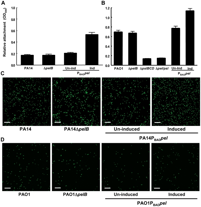
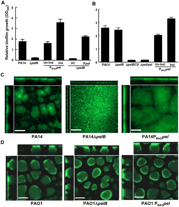
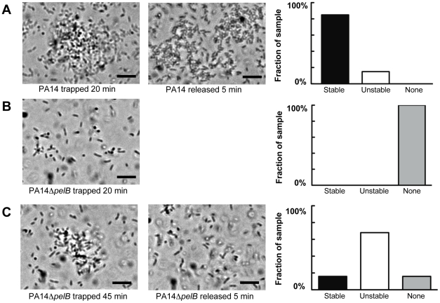
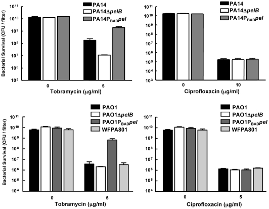
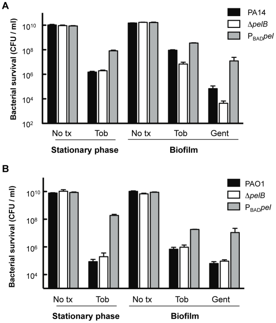
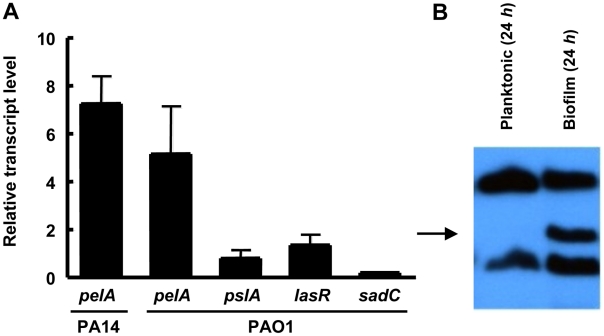
Bacterial extracellular polysaccharides are a key constituent of the extracellular matrix material of biofilms. Pseudomonas aeruginosa is a model organism for biofilm studies and produces three extracellular polysaccharides that have been implicated in biofilm development, alginate, Psl and Pel. Significant work has been conducted on the roles of alginate and Psl in biofilm development, however we know little regarding Pel. In this study, we demonstrate that Pel can serve two functions in biofilms. Using a novel assay involving optical tweezers, we demonstrate that Pel is crucial for maintaining cell-to-cell interactions in a PA14 biofilm, serving as a primary structural scaffold for the community. Deletion of pelB resulted in a severe biofilm deficiency. Interestingly, this effect is strain-specific. Loss of Pel production in the laboratory strain PAO1 resulted in no difference in attachment or biofilm development; instead Psl proved to be the primary structural polysaccharide for biofilm maturity. Furthermore, we demonstrate that Pel plays a second role by enhancing resistance to aminoglycoside antibiotics. This protection occurs only in biofilm populations. We show that expression of the pel gene cluster and PelF protein levels are enhanced during biofilm growth compared to liquid cultures. Thus, we propose that Pel is capable of playing both a structural and a protective role in P. aeruginosa biofilms.


Conflict of interest statement

The authors have declared that no competing interests exist.

Figures

Figure 1
Figure 1. Pel is not required for attachment.
Attachment of P. aeruginosa PA14 (A) and PAO1 (B) to microtiter dish wells was measured by crystal violet binding at an OD595. Overexpressing pel by addition of arabinose increased crystal violet binding in both PA14 and PAO1, but a pelB mutant showed no defect. Attachment to glass slides was visualized in a flow cell after one hour of attachment and one hour of continuous flow. Medium was supplemented with 0.2% arabinose under inducing conditions. Representative SCLM images of PA14, PA14ΔpelB and PA14PBADpel are shown using a 40× objective (C). Representative SCLM images of PAO1, PAO1ΔpelB and PAO1PBADpel are shown using a 40× objective (D). Scale bars represent 25 µm. Error bars represent standard deviations.
Figure 2
Figure 2. PA14ΔpelB is arrested in the monolayer stage of biofilm development.
Strains PA14 (A) and PAO1 (B) were compared at 24 h for levels of biomass in microtiter dishes grown at room temperature by measuring crystal violet binding OD595. Expression of the pel operon from an arabinose-inducible plasmid pMJT1 (Ppel) but not in the vector control (VC) alleviated the crystal violet staining defect of PA14ΔpelB. Biofilm structure was visualized in a flow cell and representative top-down and side-view images are shown for PA14, PA14ΔpelB and PA14PBADpel (C) and PAO1, PAO1ΔpelB and PAO1PBADpel (D). Images were obtained using a 20× objective after four d of growth in continuous flow chambers. Scale bars represent 100 µm. Error bars represent standard deviations.
Figure 3
Figure 3. Continuous Pel production is required for biofilm growth, not maintenance of existing biofilm structure.
PA14PBAD pel was grown in 2% TSB for two days under inducing conditions (0.2% arabiniose). Biofilm growth was continued either in the presence (left) or absence (right) of the inducer arabinose. Representative top-down and side-view SCLM images from day two and day four are shown. Scale bars represent 100 µm.
Figure 4
Figure 4. Pel impacts daughter cell behavior in early PA14 biofilms.
Bacterial cell divisions were monitored by time-lapse microscopy in an early-stage biofilm grown in a flow cell. Daughter cells that remained within a 15 µm diameter of the mother cell are referred to as “aggregate builders”, other cells were termed “flyers”. A minimum of 75 cell divisions was assessed for each strain.
Figure 5
Figure 5. Pel is important for cell-to-cell interactions necessary for aggregate formation.
Laser tweezers were used to trap bacteria and investigate bacterial clumping phenotypes. The captured bacteria were examined visually by light microscopy for aggregation after 20 min (A) PA14 (B) PA14ΔpelB. An extended trapping time of 45 min was required to initiate aggregation in PA14ΔpelB (C). The stability of formed aggregates was visually assessed five min after the release of the laser trap (center panel A and C). Aggregate stability was classified into three categories, “stable” if the aggregate remained intact, “unstable” if the aggregate dispersed into single cells and “none” if an aggregate did not form during the allotted time (right panels). A minimum of six replicates for each strain was assessed. Scale bars represent 10 µm. Representative phase-contrast images are shown.
Figure 6
Figure 6. Analysis of Pel-mediated antibiotic tolerance in biofilms.
48-h filter biofilms were assessed for relative susceptibility. Biofilms were treated with tobramycin and ciprofloxacin for 24 h. No antibiotic controls are included for baseline comparison. WFPA801 over-expresses the Psl polysaccharide. Bacterial survival was measured by CFU counts.
Figure 7
Figure 7. Analysis of Pel-mediated antibiotic resistance to stationary phase planktonic and biofilm grown cells.
Bacterial survival was assessed for both PA14 (A) and PAO1 (B) 24 h stationary phase cultures and 24 h filter biofilms. Prior to antibiotic treatment, stationary phase planktonic cells were centrifuged and resuspended in fresh media containing no treatment (No tx) or indicated antibiotics. Biofilm cells were moved to a fresh media source containing no treatment (No tx) or antibiotics. Planktonic cultures were treated with 5 µg/ml tobramycin (Tob). Biofilm cells were treated with either 10 µg/ml of gentamicin (Gent) or tobramycin at 5 µg/ml for PAO1 and 150 µg/ml for PA14.
Figure 8
Figure 8. pel expression is elevated during biofilm growth.
(A) Planktonic and biofilm cells are compared for transcript level by quantitative RT-PCR. In both conditions, bacteria are grown to log phase at 37°C. Planktonic cells are incubated statically in a test tube at room temperature for 30 min. Biofilm cells are grown in a tube biofilm, with an initial attachment period of 30 min followed by continuous flow for 48 h. Transcripts are normalized to ampR and then to the initial planktonic condition. Results shown are the mean of three independent experiments. Error bars represent the standard deviations. (B) Planktonic and biofilm cells were probed for PelF protein expression by western blot. A 24 h shaking liquid culture was compared to a 24 h grown tube biofilm at RT. Samples are normalized to total protein. The arrow indicates PelF expected protein size, 56 kDa.

References

    1. O'Toole GA. To build a biofilm. J Bacteriol. 2003;185:2687–2689. - PMC - PubMed
    1. Parsek MR, Singh PK. Bacterial biofilms: an emerging link to disease pathogenesis. Annu Rev Microbiol. 2003;57:677–701. - PubMed
    1. Stoodley P, Sauer K, Davies DG, Costerton JW. Biofilms as complex differentiated communities. Annu Rev Microbiol. 2002;56:187–209. - PubMed
    1. Drenkard E. Antimicrobial resistance of Pseudomonas aeruginosa biofilms. Microbes Infect. 2003;5:1213–1219. - PubMed
    1. Mah TF, O'Toole GA. Mechanisms of biofilm resistance to antimicrobial agents. Trends Microbiol. 2001;9:34–39. - PubMed

Publication types

MeSH terms