. 2011 Jan 27;6(1):e16058.
doi: 10.1371/journal.pone.0016058.

Platelet activating factor blocks interkinetic nuclear migration in retinal progenitors through an arrest of the cell cycle at the S/G2 transition

Affiliations

Platelet activating factor blocks interkinetic nuclear migration in retinal progenitors through an arrest of the cell cycle at the S/G2 transition

Lucianne Fragel-Madeira et al. PLoS One. .

Abstract

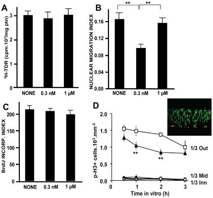
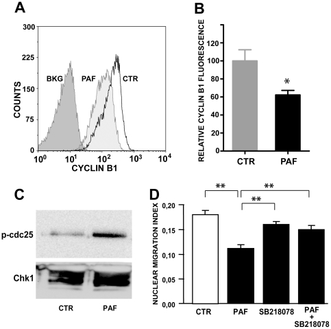
Nuclear migration is regulated by the LIS1 protein, which is the regulatory subunit of platelet activating factor (PAF) acetyl-hydrolase, an enzyme complex that inactivates the lipid mediator PAF. Among other functions, PAF modulates cell proliferation, but its effects upon mechanisms of the cell cycle are unknown. Here we show that PAF inhibited interkinetic nuclear migration (IKNM) in retinal proliferating progenitors. The lipid did not, however, affect the velocity of nuclear migration in cells that escaped IKNM blockade. The effect depended on the PAF receptor, Erk and p38 pathways and Chk1. PAF induced no cell death, nor a reduction in nucleotide incorporation, which rules out an intra-S checkpoint. Notwithstanding, the expected increase in cyclin B1 content during G2-phase was prevented in the proliferating cells. We conclude that PAF blocks interkinetic nuclear migration in retinal progenitor cells through an unusual arrest of the cell cycle at the transition from S to G2 phases. These data suggest the operation, in the developing retina, of a checkpoint that monitors the transition from S to G2 phases of the cell cycle.


Conflict of interest statement

Competing Interests: The authors have declared that no competing interests exist.

Figures

Figure 1
Figure 1. PAF and PAF receptor in the retina of developing rats.
A: Washed rabbit platelets were treated with two independent samples of retina-derived PAF-like. Positive controls were commercial PAF (10 nM) and thrombin (50 nM), WEB2086 was used at 1 µM, and silica scraped from remote areas of the TLC plate was used as negative control. Note that the PAF receptor antagonist blocked platelet aggregation induced by retina-derived PAF-like, but not by thrombin; B: PAF-like subject to reverse phase HPLC eluted as a single peak (continuous line), coincident with pure commercial PAF (dotted line); C: Western blot of PAFR among a total protein extract from P2 rat retina shows a single band. D–F: Immunohistochemical detection of PAF receptor in transverse sections of newborn rat eyes. A negative control without primary antibody is shown in F. Arrows in D show labeling consistent with a membrane receptor in various retinal layers; Arrows in E, F show optic nerve axons. GCL  =  ganglion cell layer; IPL  =  inner plexiform layer; INL  =  inner nuclear layer; NBL  =  neuroblastic layer. Scale bars in D = 50 µm; E, F = 100 µm.
Figure 2
Figure 2. PAF blocks interkinetic nuclear migration.
A–C: Sections of retinal explants immediately after dissection (A), at 3 h of incubation either in control medium (B), or with 0.3 nM PAF (C); D: A schematic diagram of interkinetic nuclear migration in the developing retina. Red elements mark the period of the cell cycle examined in the current study. E: Nuclear migration index in explants treated for 3 h with PAF. Notice the partial blockade of interkinetic nuclear migration in the range 10−10-10−8M. *  =  p<0.01 vs. control; F: BrdU incorporation index, showing a constant number of labeled nuclei across the NBL, irrespective of treatment with PAF; Data are means ± S.E.M., n = 3 in duplicate. *  =  p<0.01; **  =  p<0.001. Abbreviations as in Fig. 1.
Figure 3
Figure 3. PAF-induced blockade of nuclear migration is receptor-mediated and modulated by several signal transduction pathways.
P2 retinal explants, pre-labeled with BrdU, were treated with 0.3 nM PAF for 3 h in the presence of various PAF receptor (PAFR) antagonists or signaling inhibitors. A: Both a PAFR-ligand inactive PAF metabolite (Lyso-PAF 10 nM, left) and a PAFR antagonist (WEB2086 10 nM, right) prevented the effect of PAF; B: Deletion of PAFR abrogates the effect of PAF upon interkinetic nuclear migration; C–E: Antagonists of both Erk (C) and p38 (D) MAP kinases prevented the effect of PAF, whereas an inhibitor of protein kinase C (E) had no effect. Data are means ± S.E.M., n = 4 in duplicate. **  =  p<0.001.
Figure 4
Figure 4. Nuclei of proliferating cells at the end of the S-phase escape PAF-induced blockade, and PAF does not interfere with nuclear movement proper.
A–C: High magnification of immunolabeled sections of retinal explants immediately after dissection (A), at 3 h of incubation either in control medium (B), or with 1 nM PAF (C). Arrows highlight the mottled, predominantly peripheral pattern of BrdU incorporation in nuclei departing from the S-phase stratum (A), and in the reduced number of nuclei that reach the outer portions of the neuroblastic layer in PAF-treated explant (C), when compared with control (B). This labeling pattern is typical of nucleotide incorporation at the end of S-phase. Scale bar  = 10 µm. D–F: Radially cut P2 retinal explants were pulse-labeled with BrdU in vitro and treated with 0.3 nM PAF for 0.5, 1, 2 or 3 hours. Then, transverse sections immunolabeled for BrdU were used to map the location of the furthest migrating nuclei in 5 locations along the centro-peripheral extent of the retina (central = 1; peripheral = 5). The mean average distances (wavefronts) travelled by the furthest migrating nuclei are shown in D, as percentage of the basal-to-apical extent of the neuroblastic layer, as a function of retinal eccentricity from the optic disk. Open circles  =  control, filled triangles  =  PAF-treated. Note the coincident migration wavefronts, despite the distinct nuclear migration indexes (E, *  =  p<0.01 vs. control). Constant BrdU labeling index (F) is a control.
Figure 5
Figure 5. PAF induces a post-replication cell cycle arrest.
A–C: Treatment with PAF does not block DNA replication. Tritiated thymidine was equally incorporated in BrdU-labeled control and PAF-treated explants (A), irrespective of blockade of interkinetic nuclear migration (B and C are controls from the same batch of explants used for the 3H-TDR measurements); **  =  p<0.001. D: Arrested nuclei do not progress along the cell cycle. Explants from the retina of animals pre-injected with BrdU were maintained either in control medium or treated with 0.3 nM PAF for various intervals, and retinal sections were stained with antibodies to BrdU (green in the inset) and to phospho-histone H3 (red). Despite the blockade of nuclear migration, no immunolabeling was detected among nuclei arrested within the inner 2/3 of the neuroblastic layer (inset), and all pH3-labeled nuclei were located within the outer (apical) 1/3, albeit with a distinctive reduction in PAF-treated explants. Data are means ± S.E.M., n = 4 in duplicate; **  =  p<0.001 vs. control at same time in vitro.
Figure 6
Figure 6. Lack of significant DNA damage induced by PAF.
A–B: Photomicrographs of the nuclei of cells dissociated from retinal explants either untreated (A) or incubated with 0.3 nM PAF (B), double-labeled with the DNA marker SYTOX green, and with a monoclonal antibody to p-H2AX developed with a red fluorochrome-labeled secondary antibody. C: Frequency of cells showing foci of p-H2AX in cells from either untreated or PAF-treated retinal explants. Data are means ± S.E.M., pooled from 4 independent experiments.
Figure 7
Figure 7. PAF blocks cyclin B1 increase during G2 phase, and PAF-induced cell cycle arrest depends on Chk1.
A, B: Analysis of cyclin B1 content in BrdU-labeled cells. A: Cytofluorograms gated on BrdU-labeled cells dissociated from either control or PAF-treated retinal explants shows displacement of PAF-treated cells towards lower levels of cyclin B1; BKG  =  controls without primary cyclin B1 antibody; B: Mean and S.E.M. of the median cyclin B1 fluorescent intensity in BrdU-labeled cells, averaged among 4 independent experiments. Data were normalized to the respective controls in each experiment; *  =  p<0.01. C: Treatment of retinal explants with 0.3 nM PAF for 3 h induces an increase in the activity of Chk1, as shown by increased phosphorylation of a target cdc25C peptide (n = 2 with identical results) D: SB218078, a Chk1 inhibitor, blocks the effect of PAF (Data are means ± S.E.M., from n = 5 independent experiments in duplicate); **  =  p<0.001.

Similar articles

Cited by

References

    1. Boye E, Skjolberg HC, Grallert B. Checkpoint regulation of DNA replication. Methods Mol Biol. 2009;521:55–70. - PubMed
    1. Ciliberto A, Shah JV. A quantitative systems view of the spindle assembly checkpoint. EMBO J. 2009;28:2162–2173. - PMC - PubMed
    1. Chin CF, Yeong FM. Safeguarding entry into mitosis: the antephase checkpoint. Mol Cell Biol. 2010;30:22–32. - PMC - PubMed
    1. Cook JG. Replication licensing and the DNA damage checkpoint. Front Biosci. 2009;14:5013–5030. - PMC - PubMed
    1. Chow JP, Siu WY, Ho HT, Ma KH, Ho CC, et al. Differential contribution of inhibitory phosphorylation of CDC2 and CDK2 for unperturbed cell cycle control and DNA integrity checkpoints. J Biol Chem. 2003;278:40815–40828. - PubMed

Publication types

Substances