. 2011 Jan 27;6(1):e14593.
doi: 10.1371/journal.pone.0014593.

Epigenetics underpinning the regulation of the CXC (ELR+) chemokines in non-small cell lung cancer

Affiliations

Epigenetics underpinning the regulation of the CXC (ELR+) chemokines in non-small cell lung cancer

Anne-Marie Baird et al. PLoS One. .

Abstract

Background: Angiogenesis may play a role in the pathogenesis of Non-Small Cell Lung cancer (NSCLC). The CXC (ELR(+)) chemokine family are powerful promoters of the angiogenic response.

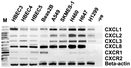
Methods: The expression of the CXC (ELR(+)) family members (CXCL1-3/GROα-γ, CXCL8/IL-8, CXCR1/2) was examined in a series of resected fresh frozen NSCLC tumours. Additionally, the expression and epigenetic regulation of these chemokines was examined in normal bronchial epithelial and NSCLC cell lines.

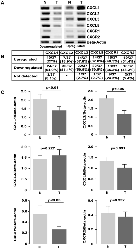
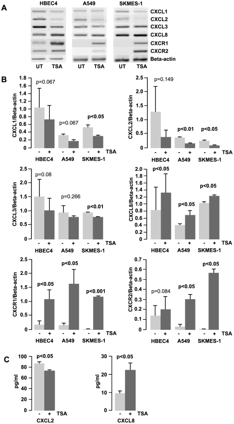
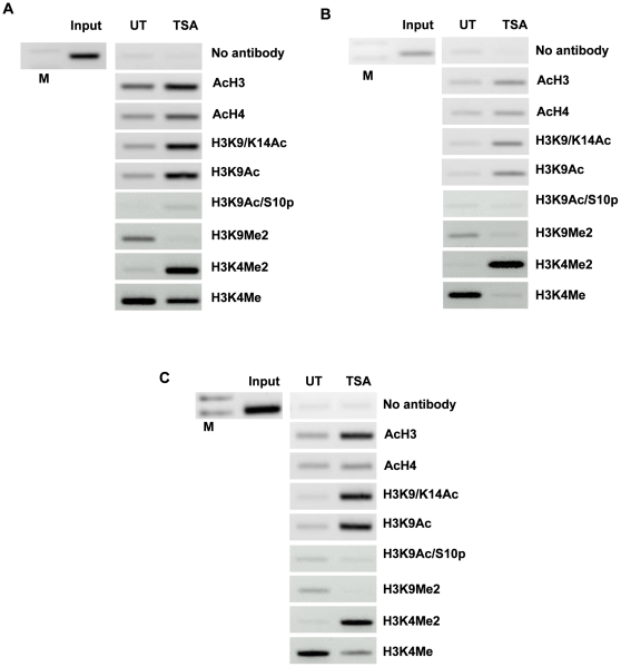
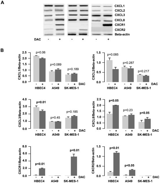
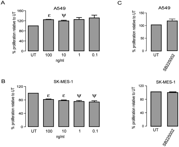
Results: Overall, expression of the chemokine ligands (CXCL1, 2, 8) and their receptors (CXCR1/2) were down regulated in tumour samples compared with normal, with the exception of CXCL3. CXCL8 and CXCR1/2 were found to be epigenetically regulated by histone post-translational modifications. Recombinant CXCL8 did not stimulate cell growth in either a normal bronchial epithelial or a squamous carcinoma cell line (SKMES-1). However, an increase was observed at 72 hours post treatment in an adenocarcinoma cell line.

Conclusions: CXC (ELR(+)) chemokines are dysregulated in NSCLC. The balance of these chemokines may be critical in the tumour microenvironment and requires further elucidation. It remains to be seen if epigenetic targeting of these pathways is a viable therapeutic option in lung cancer treatment.


Conflict of interest statement

Competing Interests: Funding for this project was provide by Pfizer in the form of a salary for Anne-Marie Baird. No funding was provided for consumables. The funding provided was a grant-in-aid, and as such, it does not alter the authors' adherence to all the PLoS ONE policies on sharing data and materials.

Figures

Figure 1
Figure 1. CXC (ELR+) chemokine family mRNA expression in normal/tumour NSCLC matched pairs.
A) Levels of CXCL13, CXCL8 and CXCR1/2 were examined by RT-PCR on a panel of NSCLC lines (adenocarcinoma (n = 14), squamous cell carcinoma (n = 23)) patient samples. Representative images of up and down regulated samples are shown. Beta actin levels were used for normalisation purposes. B) A summary of the changes in expression of the various CXC (ELR+) chemokines and their receptors in a panel of tumour/normal matched patient NSCLC samples. C) Overall densitometry analyses of tumour/normal matched patient NSCLC samples. Data is graphed as mean ± standard error of mean (n = 37). (N – Normal, T – Tumour).
Figure 2
Figure 2. Expression of CXCL13, CXCL8 and CXCR1/2 in a panel of normal and NSCLC cancer cell lines.
The panel included A549 (adenocarcinoma), SKMES-1 (squamous cell carcinoma), H460, H647 and H1299 (large cell carcinoma), BEAS-2B (SV40 transformed normal bronchoepithelial) and HBEC cell lines (normal bronchial epithelial cell lines immortalised in the absence of viral oncoproteins). Beta actin is included to validate loading efficiency. (M – DNA size marker, -ve – Negative RT-PCR control).
Figure 3
Figure 3. Cell line response to histone deacetylase inhibition.
A) The effect of TSA treatment (250 ng/mL for 16 h) on the expression of CXCL13, CXCL8 and CXCR1/2. B) Densitometry analysis of expression in treated versus untreated samples when normalised to beta actin. Data is graphed as mean ± standard error of mean (n = 3). C) Treatment with TSA also effects the production of CXCL2 and CXCL8 at protein level in SKMES-1 cells. Chemokines were quantified in conditioned media removed from culture after exposure to TSA (250 ng/mL for 16 h). Data is graphed as mean ± standard error of mean (n = 3). (UT – untreated, TSA – Trichostatin A).
Figure 4
Figure 4. Histone acetylation occurs directly at the promoter regions of CXCL8 and CXCR1/2.
The ChIP assay demonstrates that TSA treatment results in an increase in the acetylation of histone H3 and H4. A549 cells were cultured in the presence or absence of TSA (250 ng/mL) for a period of 16 h. Subsequently, a ChIP assay was performed using the following antibodies; pan acetylated histone H3 (Ac H3) and H4 (Ac H4), histone H3 acetylated at lysine 9 and 14 (H3K9/K14Ac), histone H3 acetylated at lysine 9 (H3K9Ac), histone H3 acetylated at lysine 9 and phosphorylated at serine 10 (H3K9pS10), Histone H3 dimetylation marker at lysine 9 (H3K9Me2), dimetylation marker at lysine 4 (H3K4Me2) and methylation marker at lysine 4 (H3K4Me). The chromatin status at the promoter region of (A) CXCL8, (B) CXCR1 and (C) CXCR2 is shown. Input DNA serves as a positive control recommended by the manufacturer (Diagenode). A no antibody control was included to test for non specific carriage of DNA with histones. (M – DNA size ladder).
Figure 5
Figure 5. Cell line response to a DNA methyltransferase inhibitor.
A) The effect of 5-aza-2′deoxycytidine (DAC) treatment on the expression of CXCL13, CXCL8 and CXCR1/2. Cells were cultured in 1 µM DAC for 48 h with media and drug replaced every 24 h. Expression changes were measured using RT-PCR with Beta-Actin used as the internal control for quantification purposes. B) Densitometry analysis of expression in treated versus untreated samples when normalised to beta actin. Data is graphed as mean ± standard error of mean. (n = 3). (DAC – 5-aza-2′deoxycitidine).
Figure 6
Figure 6. The growth effect of CXCL8 varies between NSCLC sub-type but is negated by neutralizing the CXCR2 receptor.
A) Cell proliferation was examined by BrdU assay following 24–72 h treatment with CXCL8 in A549 (72 h post treatment) and SKMES-1 (24 h post treatment) cell lines. Only time points with significant data is shown. Data is represented as a percentage of the untreated control (UT), which was set to 100% and is expressed as mean ± SEM. (n = 3) (ε p<0.01 – CXCL8 treatment vs. UT, ψ p<0.05 CXCL8 treatment vs. UT) B) Cell lines were treated with a selective CXCR2 antagonist (0.022µM) for 1 h prior to the addition of CXCL8 and cultured for a period of 24 (SKMES-1) or 72 h (A549). Data is represented as a percentage of the untreated control (UT), which was set to 100% and is expressed as mean ± SEM. (n = 3).

References

    1. O'Byrne KJ, Dalgleish AG, Browning MJ, Steward WP, Harris AL. The relationship between angiogenesis and the immune response in carcinogenesis and the progression of malignant disease. Eur J Cancer. 2000;36:151–169. - PubMed
    1. Arenberg DA, Polverini PJ, Kunkel SL, Shanafelt A, Hesselgesser J, et al. The role of CXC chemokines in the regulation of angiogenesis in non-small cell lung cancer. J Leukoc Biol. 1997;62:554–562. - PubMed
    1. Zlotnik A, Yoshie O. Chemokines: a new classification system and their role in immunity. Immunity. 2000;12:121–127. - PubMed
    1. Strieter RM, Burdick MD, Gomperts BN, Belperio JA, Keane MP. CXC chemokines in angiogenesis. Cytokine Growth Factor Rev. 2005;16:593–609. - PubMed
    1. Addison CL, Daniel TO, Burdick MD, Liu H, Ehlert JE, et al. The CXC chemokine receptor 2, CXCR2, is the putative receptor for ELR+ CXC chemokine-induced angiogenic activity. J Immunol. 2000;165:5269–5277. - PubMed

Publication types

MeSH terms