

Microarray analysis on human neuroblastoma cells exposed to aluminum, β(1-42)-amyloid or the β(1-42)-amyloid aluminum complex
- PMID: 21298039
- PMCID: PMC3029275
- DOI: 10.1371/journal.pone.0015965
Microarray analysis on human neuroblastoma cells exposed to aluminum, β(1-42)-amyloid or the β(1-42)-amyloid aluminum complex
Abstract
Background: A typical pathological feature of Alzheimer's disease (AD) is the appearance in the brain of senile plaques made up of β-amyloid (Aβ) and neurofibrillary tangles. AD is also associated with an abnormal accumulation of some metal ions, and we have recently shown that one of these, aluminum (Al), plays a relevant role in affecting Aβ aggregation and neurotoxicity.
Methodology: In this study, employing a microarray analysis of 35,129 genes, we investigated the effects induced by the exposure to the Aβ(1-42)-Al (Aβ-Al) complex on the gene expression profile of the neuronal-like cell line, SH-SY5Y.
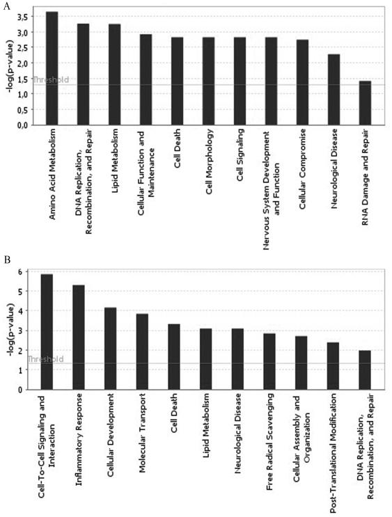
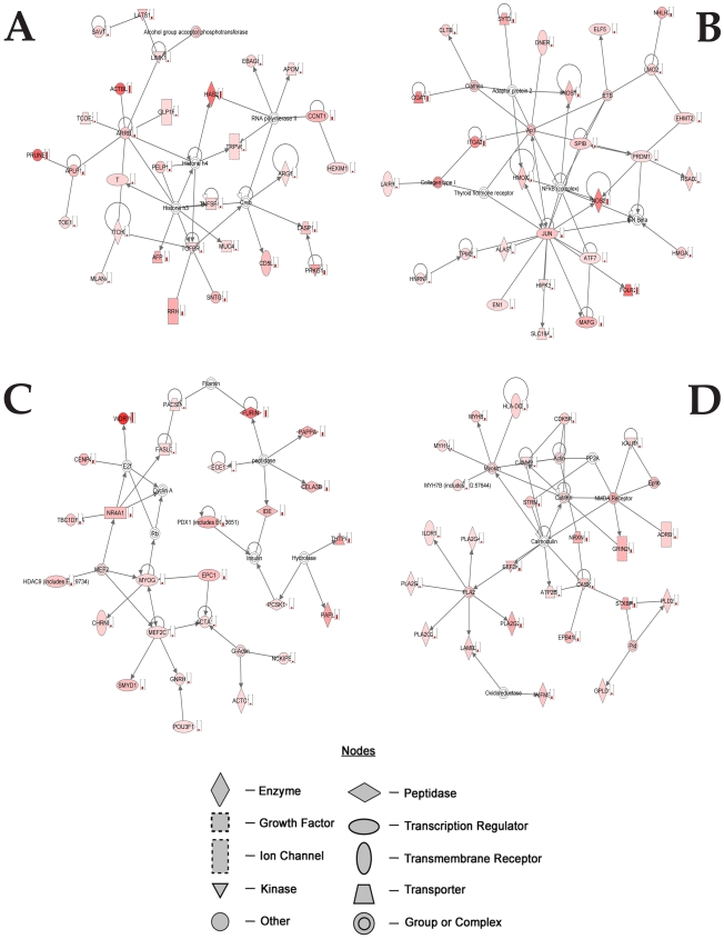
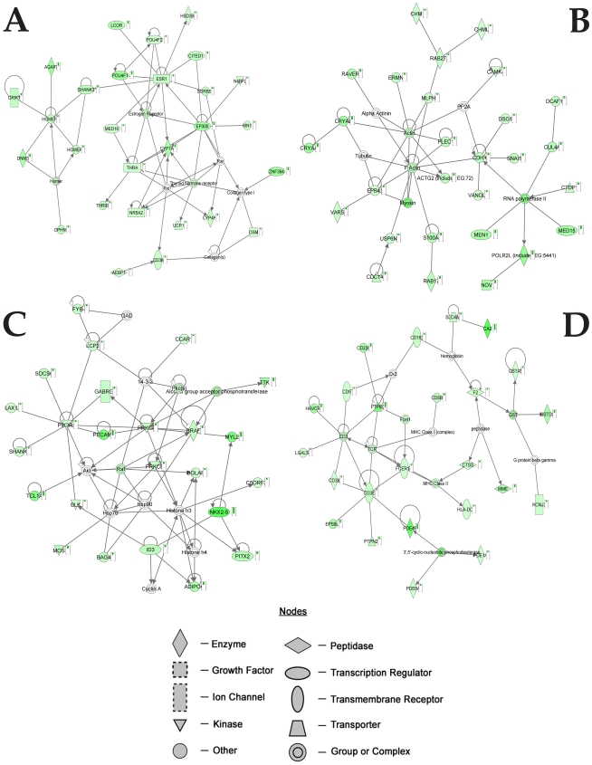
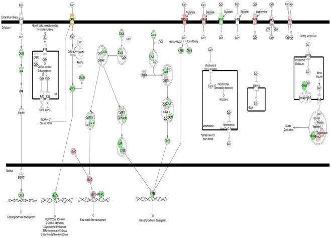
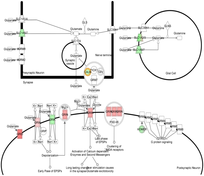
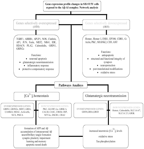
Principal findings: The microarray assay indicated that, compared to Aβ or Al alone, exposure to Aβ-Al complex produced selective changes in gene expression. Some of the genes selectively over or underexpressed are directly related to AD. A further evaluation performed with Ingenuity Pathway analysis revealed that these genes are nodes of networks and pathways that are involved in the modulation of Ca(2+) homeostasis as well as in the regulation of glutamatergic transmission and synaptic plasticity.
Conclusions and significance: Aβ-Al appears to be largely involved in the molecular machinery that regulates neuronal as well as synaptic dysfunction and loss. Aβ-Al seems critical in modulating key AD-related pathways such as glutamatergic transmission, Ca(2+) homeostasis, oxidative stress, inflammation, and neuronal apoptosis.
Conflict of interest statement
Figures
References
-
- Lovell MA, Robertson JD, Teesdale WJ, Campbell JL, Markesbery WR. Copper, iron and zinc in Alzheimer's disease senile plaques. J Neurol Sci. 1998;158:47–52. - PubMed
-
- Good PF, Perl DP, Bierer LM, Schmeidler J. Selective accumulation of aluminum and iron in the neurofibrillary tangles of Alzheimer's disease: a laser microprobe (LAMMA) study. Ann Neurol. 1992;31:286–292. - PubMed
-
- Liu G, Huang W, Moir RD, Vanderburg CR, Lai B, et al. Metal exposure and Alzheimer's pathogenesis. J Struct Biol. 2006;155:45–51. - PubMed
-
- Sensi SL, Paoletti P, Bush AI, Sekler I. Zinc in the physiology and pathology of the CNS. Nat Rev Neurosci. 2009;10:780–791. - PubMed
-
- House E, Collingwood J, Khan A, Korchazkina O, Berthon G, et al. Aluminium, iron, zinc and copper influence the in vitro formation of amyloid fibrils of Abeta42 in a manner which may have consequences for metal chelation therapy in Alzheimer's disease. J Alzheimers Dis. 2004;6:291–301. - PubMed
Publication types
MeSH terms
Substances
LinkOut - more resources
Full Text Sources
Medical
Molecular Biology Databases
Miscellaneous
