

Extrasynaptic GABA(A) receptors and tonic inhibition in rat auditory thalamus
- PMID: 21298071
- PMCID: PMC3027696
- DOI: 10.1371/journal.pone.0016508
Extrasynaptic GABA(A) receptors and tonic inhibition in rat auditory thalamus
Abstract
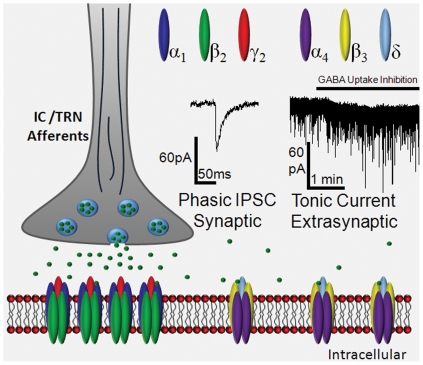
Background: Neural inhibition plays an important role in auditory processing and attentional gating. Extrasynaptic GABA(A) receptors (GABA(A)R), containing α(4)and δ GABA(A)R subunits, are thought to be activated by GABA spillover outside of the synapse following release resulting in a tonic inhibitory Cl(-) current which could account for up to 90% of total inhibition in visual and somatosensory thalamus. However, the presence of this unique type of inhibition has not been identified in auditory thalamus.
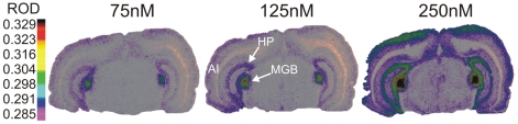
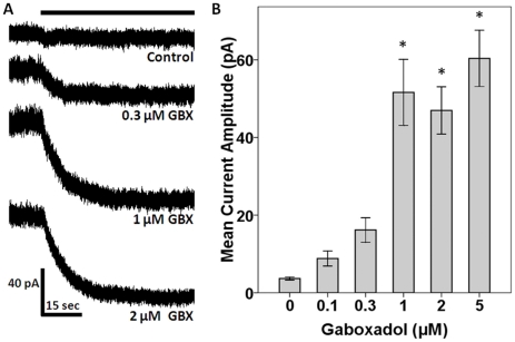
Methodology/principal findings: The present study used gaboxadol, a partially selective potent agonist for δ-subunit containing GABA(A) receptor constructs to elucidate the presence of extrasynaptic GABA(A)Rs using both a quantitative receptor binding assay and patch-clamp electrophysiology in thalamic brain slices. Intense [(3)H]gaboxadol binding was found to be localized to the MGB while whole cell recordings from MGB neurons in the presence of gaboxadol demonstrated the expression of δ-subunit containing GABA(A)Rs capable of mediating a tonic inhibitory Cl(-) current.
Conclusions/significance: Potent tonic inhibitory GABA(A)R responses mediated by extrasynaptic receptors may be important in understanding how acoustic information is processed by auditory thalamic neurons as it ascends to auditory cortex. In addition to affecting cellular behavior and possibly neurotransmission, functional extrasynaptic δ-subunit containing GABA(A)Rs may represent a novel pharmacological target for the treatment of auditory pathologies including temporal processing disorders or tinnitus.
Conflict of interest statement
Figures
Similar articles
-
Reduced GABA(A) receptor-mediated tonic inhibition in aged rat auditory thalamus.J Neurosci. 2013 Jan 16;33(3):1218-27a. doi: 10.1523/JNEUROSCI.3277-12.2013. J Neurosci. 2013. PMID: 23325258 Free PMC article.
-
An extrasynaptic GABAA receptor mediates tonic inhibition in thalamic VB neurons.J Neurophysiol. 2005 Dec;94(6):4491-501. doi: 10.1152/jn.00421.2005. Epub 2005 Sep 14. J Neurophysiol. 2005. PMID: 16162835
-
Is GABA neurotransmission enhanced in auditory thalamus relative to inferior colliculus?J Neurophysiol. 2014 Jan;111(2):229-38. doi: 10.1152/jn.00556.2013. Epub 2013 Oct 23. J Neurophysiol. 2014. PMID: 24155003 Free PMC article.
-
Auditory thalamic circuits and GABAA receptor function: Putative mechanisms in tinnitus pathology.Hear Res. 2017 Jun;349:197-207. doi: 10.1016/j.heares.2016.08.009. Epub 2016 Aug 21. Hear Res. 2017. PMID: 27553899 Free PMC article. Review.
-
GABAA receptors in the thalamus: alpha4 subunit expression and alcohol sensitivity.Alcohol. 2007 May;41(3):177-85. doi: 10.1016/j.alcohol.2007.03.010. Epub 2007 May 23. Alcohol. 2007. PMID: 17521848 Review.
Cited by
-
GABAergic inhibition shapes SAM responses in rat auditory thalamus.Neuroscience. 2015 Jul 23;299:146-55. doi: 10.1016/j.neuroscience.2015.04.062. Epub 2015 May 2. Neuroscience. 2015. PMID: 25943479 Free PMC article.
-
The Cerebellar GABAAR System as a Potential Target for Treating Alcohol Use Disorder.Handb Exp Pharmacol. 2018;248:113-156. doi: 10.1007/164_2018_109. Handb Exp Pharmacol. 2018. PMID: 29736774 Free PMC article.
-
Impact of ageing on postsynaptic neuronal nicotinic neurotransmission in auditory thalamus.J Physiol. 2017 Aug 1;595(15):5375-5385. doi: 10.1113/JP274467. Epub 2017 Jul 7. J Physiol. 2017. PMID: 28585699 Free PMC article.
-
The FBN rat model of aging: investigation of ABR waveforms and ribbon synapse changes.Neurobiol Aging. 2018 Feb;62:53-63. doi: 10.1016/j.neurobiolaging.2017.09.034. Epub 2017 Oct 9. Neurobiol Aging. 2018. PMID: 29107847 Free PMC article.
-
Effect of auditory cortex deactivation on stimulus-specific adaptation in the medial geniculate body.J Neurosci. 2011 Nov 23;31(47):17306-16. doi: 10.1523/JNEUROSCI.1915-11.2011. J Neurosci. 2011. PMID: 22114297 Free PMC article.
References
-
- Yu XJ, Xu XX, He S, He J. Change detection by thalamic reticular neurons. NatNeurosci. 2009;12:1165–1170. - PubMed
-
- Bartlett EL, Wang X. Neural representations of temporally modulated signals in the auditory thalamus of awake primates. JNeurophysiol. 2007;97:1005–1017. - PubMed
-
- Paxinos W, Watson C. San Diego: Academic Press; 1998. The Rat Brain in Stereotaxic Coordinates.
-
- Clerici WJ, Coleman JR. Postnatal cytoarchitecture of the rat medial geniculate body. JComp Neurol. 1998;399:110–124. - PubMed
Publication types
MeSH terms
Substances
Grants and funding
LinkOut - more resources
Full Text Sources
Other Literature Sources
