. 2011 Jan 27;7(1):e1001285.
doi: 10.1371/journal.pgen.1001285.

Evolutionary conserved regulation of HIF-1β by NF-κB

Affiliations

Evolutionary conserved regulation of HIF-1β by NF-κB

Patrick van Uden et al. PLoS Genet. .

Abstract

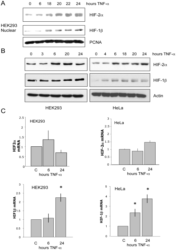
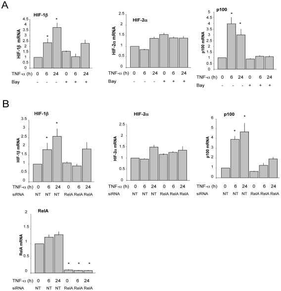
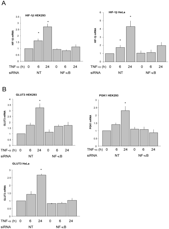
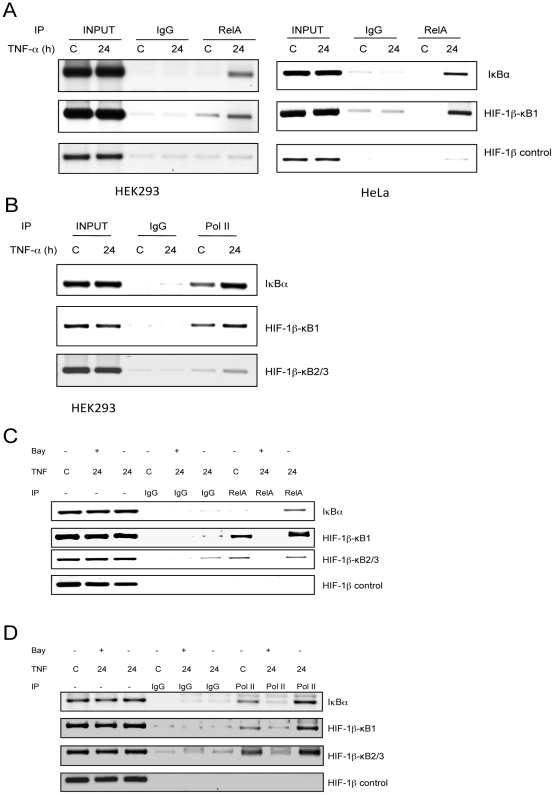
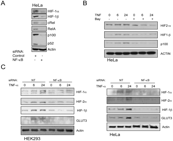
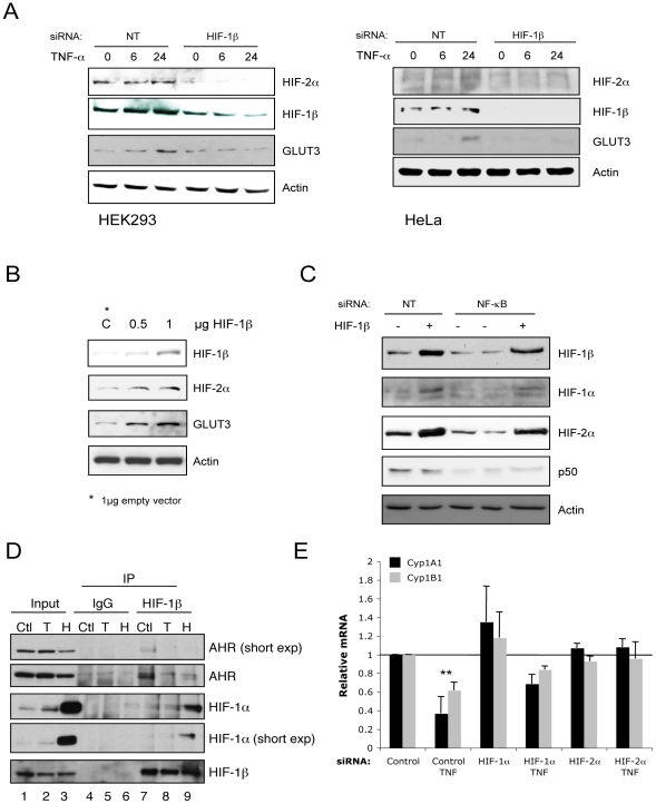
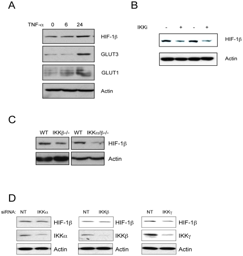
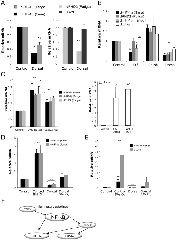
Hypoxia Inducible Factor-1 (HIF-1) is essential for mammalian development and is the principal transcription factor activated by low oxygen tensions. HIF-α subunit quantities and their associated activity are regulated in a post-translational manner, through the concerted action of a class of enzymes called Prolyl Hydroxylases (PHDs) and Factor Inhibiting HIF (FIH) respectively. However, alternative modes of HIF-α regulation such as translation or transcription are under-investigated, and their importance has not been firmly established. Here, we demonstrate that NF-κB regulates the HIF pathway in a significant and evolutionary conserved manner. We demonstrate that NF-κB directly regulates HIF-1β mRNA and protein. In addition, we found that NF-κB-mediated changes in HIF-1β result in modulation of HIF-2α protein. HIF-1β overexpression can rescue HIF-2α protein levels following NF-κB depletion. Significantly, NF-κB regulates HIF-1β (tango) and HIF-α (sima) levels and activity (Hph/fatiga, ImpL3/ldha) in Drosophila, both in normoxia and hypoxia, indicating an evolutionary conserved mode of regulation. These results reveal a novel mechanism of HIF regulation, with impact in the development of novel therapeutic strategies for HIF-related pathologies including ageing, ischemia, and cancer.


Conflict of interest statement

The authors have declared that no competing interests exist.

Figures

Figure 1
Figure 1. TNF-α induces HIF-1β and HIF-2α protein but only HIF-1β mRNA.
(A) HEK293 were treated with 20 ng/mL TNF-α for the indicated times prior to nuclear extraction. Levels of HIF-2α and HIF-1b were analysed by Western Blot. PCNA was used as a loading control. (B) HEK293 and HeLa cells were treated as in A, prior to lysis. Whole cell lysates were analysed by Western Blot. (C) HEK293 and HeLa cells were treated with 20 ng/mL TNF-α for the indicated periods of times prior to total RNA extraction. Following cDNA synthesis, qPCR was performed for the levels of HIF-2α and HIF-1β.
Figure 2
Figure 2. TNF-α–induced HIF-1β mRNA is IKK- and RelA-dependent.
(A) HEK293 cells were pre-treated with the IKK inhibitor Bay 11 7082 prior to treatment with 20 ng/mL TNF-α for the indicated times. Total RNA was extracted, converted to cDNA, and qPCR was performed for the levels of HIF-1β, HIF-2α and p100. (B) HEK293 cells were transfected with control and RelA siRNA oligonucleotides for a total of 72 hours. Where indicated cells were treated with 20 ng/mL TNF-α prior to total RNA extraction. qPCR was performed for the levels of HIF-1β, HIF-2α, p100 and RelA.
Figure 3
Figure 3. TNF-α–induced HIF mRNA levels and activity are NF-κB–dependent.
(A) HEK293 and HeLa cells were transfected with control and pan-NF-κB siRNA oligonucleotides for a total of 72 hours. Where indicated cells were treated with 20 ng/mL TNF-α prior to total RNA extraction. qPCR was performed for the levels of HIF-1β. (B) Cells were treated and processed as in A, and qPCR was performed for the indicated genes.
Figure 4
Figure 4. TNF-α induces RelA and RNA polymerase II recruitment to the HIF-1β promoter.
(A) HEK293 and HeLa cells were treated with 20 ng/mL TNF-α for 24 hours prior to crosslinking and lysis. ChIPs were performed using the indicated antibodies and purified DNA was amplified using PCR primers for the HIF-1β promoter and control regions. The IκB-α promoter was used as a positive control. (B) ChIPs were performed for RNA polymerase II and purified DNA processed as in A. (C) HeLa cells were pre-treated with the IKK inhibitor Bay 11 7082 for 30 minutes prior to TNF-α treatment. Cells were crosslinked and lysed 24 hours later. ChIPs were performed for the levels of RelA. (D) ChIPs were performed for RNA polymerase II and purified DNA processed as in A. IκB-α promoter was used as a positive control.
Figure 5
Figure 5. TNF-α–induced HIF-1β and HIF-2α proteins are IKK- and NF-κB–dependent.
(A) HeLa cells were transfected with control or pan-NF-κB siRNA oligonucleotides and cultured for a total of 48 hours. Whole cell lysates were analysed by Western blot for the proteins mentioned. (B) HeLa cells were pre-treated with the IKK inhibitor Bay 11 7082 for 30 minutes prior to TNF-α treatment for the indicated periods of time prior to lysis. Whole cell lysates were analysed by Western blot for the proteins mentioned. (C) HEK293 and HeLa cells were transfected with control or pan-NF-κB siRNA oligonucleotides and cultured for a total of 72 hours. Where indicated, cells were treated with 20 ng/mL TNF-α prior to lysis. Whole cell lysates were analysed by Western blot for the proteins mentioned.
Figure 6
Figure 6. HIF-1β is required for TNF-α–induced HIF-2α, which represses AHR function.
(A) HEK293 and HeLa cells were transfected with control or HIF-1β siRNA oligonucleotides and cultured for a total of 72 hours. Where indicated cells were treated with 20 ng/mL TNF-α prior to lysis. Whole cell lysates were analysed by Western blot for the proteins mentioned. (B) HEK293 cells were transfected with increasing amount of HIF-1β expression plasmid for 48 hours prior to lysates. Whole cell lysates were analysed by Western blot for the indicated proteins. (C) HEK293 cells were transfected with control or pan-NF-κB siRNA oligonucleotides and cultured for 72 hours. Where indicated cells were co-transfected with HIF-1β expression plasmids. Whole cell lysates were analysed by Western blot for the mentioned proteins. (D) HeLa cells were left untreated (Ctl) treated with 20 ng/mL TNF-α (T) or 1% O2 (H) for 24 hours prior to lysis. 200 µg of protein were used to immunoprecipitate HIF-1β. Levels of associated proteins were assessed by western blot using the indicated antibodies. Input corresponds to 10% of starting material. (E) HeLa cells were transfected with control, HIF-1α or HIF-2α siRNA oligonucleotides for a total of 48 hours. Where indicated cells were treated with 20 ng/mL TNF-α prior to total RNA extraction. qPCR was performed for the levels of the indicated genes.
Figure 7
Figure 7. NF-κB–mediated control of the HIF system is conserved in mice.
(A) Mouse embryo fibroblasts (MEFs) were treated with 20 ng/mL TNF-α for the indicated period of time prior to lysis. Whole cell lysates were analysed by Western blot. (B) MEFs were treated with the IKK inhibitor for 24 hours prior to lysis. Whole cell lysates were analysed by Western blot for the levels of HIF-1β. (C) Whole cell lysates were obtained from IKK wildtype (WT), IKKβ-/- or IKKα/β-/- MEFs and analysed by western blots. (D) MEFs were transfected with siRNA oligonucleotides for IKKα, IKKβ or IKKγ and the levels of HIF-1β were analysed by western blot.
Figure 8
Figure 8. NF-κB–mediated control of the HIF system is conserved in Drosophila.
(A) Total RNA was extracted from control and Dorsal null flies, and qPCR was performed for the levels of Drosophila HIF-1α, HIF-1β, PHD and ISWI. (B) Total RNA was extracted from control Dif, Relish and Dorsal null flies, and qPCR was performed for the levels of the indicated genes. (C). Total RNA was extracted from control, UAS-Dorsal or Cactus null flies, and qPCR was performed for the levels of the indicated genes. (D) Total RNA was extracted from control and Dorsal null flies exposed or not to 5% O2 for 24 hours. qPCR was performed for the levels of Drosophila HIF-1α, HIF-1β. (E) As in D, but qPCR was performed for the levels of Drosophila PHD and Ldha. (F) Proposed model of NF-κB modulation of the HIF pathway. NF-κB directly regulates HIF-1α and HIF-1β genes. NF-κB induced HIF-1β mediates HIF-2α stability.

Similar articles

Cited by

References

    1. Rocha S. Gene regulation under low oxygen: holding your breath for transcription. Trends Biochem Sci. 2007;32:389–397. - PubMed
    1. Kenneth NS, Rocha S. Regulation of gene expression by hypoxia. Biochem J. 2008;414:19–29. - PubMed
    1. Fandrey J, Gorr TA, Gassmann M. Regulating cellular oxygen sensing by hydroxylation. Cardiovasc Res. 2006;71:642–651. - PubMed
    1. Gorr TA, Gassmann M, Wappner P. Sensing and responding to hypoxia via HIF in model invertebrates. J Insect Physiol. 2006;52:349–364. - PubMed
    1. Epstein AC, Gleadle JM, McNeill LA, Hewitson KS, O'Rourke J, et al. C. elegans EGL-9 and mammalian homologs define a family of dioxygenases that regulate HIF by prolyl hydroxylation. Cell. 2001;107:43–54. - PubMed

Publication types

Substances

LinkOut - more resources