. 2011 Jan 26;6(1):e16199.
doi: 10.1371/journal.pone.0016199.

Mammalian frataxin: an essential function for cellular viability through an interaction with a preformed ISCU/NFS1/ISD11 iron-sulfur assembly complex

Affiliations

Mammalian frataxin: an essential function for cellular viability through an interaction with a preformed ISCU/NFS1/ISD11 iron-sulfur assembly complex

Stéphane Schmucker et al. PLoS One. .

Abstract

Background: Frataxin, the mitochondrial protein deficient in Friedreich ataxia, a rare autosomal recessive neurodegenerative disorder, is thought to be involved in multiple iron-dependent mitochondrial pathways. In particular, frataxin plays an important role in the formation of iron-sulfur (Fe-S) clusters biogenesis.

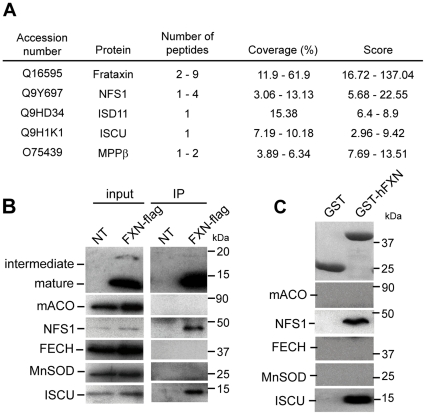
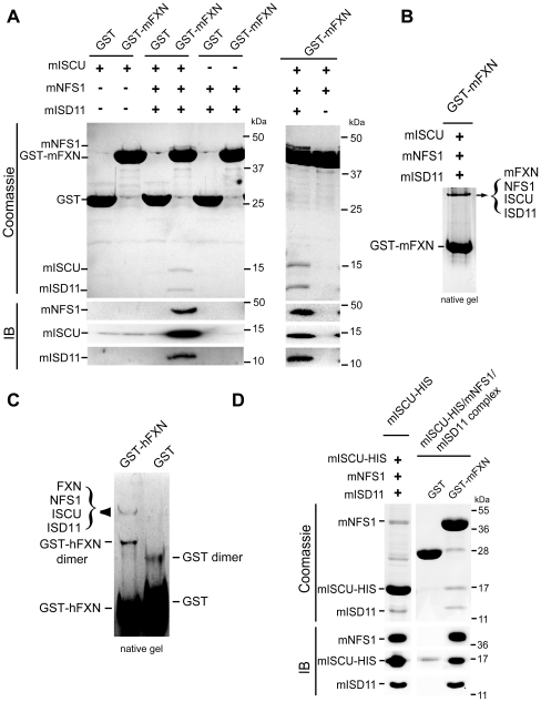
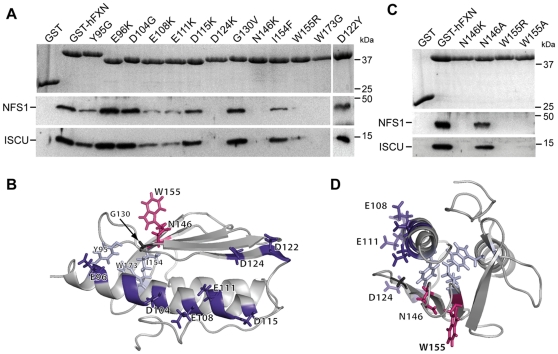
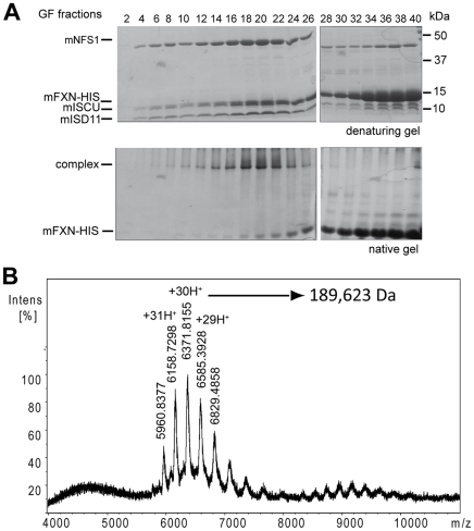
Methodology/principal findings: We present data providing new insights into the interactions of mammalian frataxin with the Fe-S assembly complex by combining in vitro and in vivo approaches. Through immunoprecipitation experiments, we show that the main endogenous interactors of a recombinant mature human frataxin are ISCU, NFS1 and ISD11, the components of the core Fe-S assembly complex. Furthermore, using a heterologous expression system, we demonstrate that mammalian frataxin interacts with the preformed core complex, rather than with the individual components. The quaternary complex can be isolated in a stable form and has a molecular mass of ≈190 kDa. Finally, we demonstrate that the mature human FXN(81-210) form of frataxin is the essential functional form in vivo.

Conclusions/significance: Our results suggest that the interaction of frataxin with the core ISCU/NFS1/ISD11 complex most likely defines the essential function of frataxin. Our results provide new elements important for further understanding the early steps of de novo Fe-S cluster biosynthesis.


Conflict of interest statement

Competing Interests: The authors have declared that no competing interests exist.

Figures

Figure 1
Figure 1. Human frataxin interacts specifically with ISCU, NFS1 and ISD11.
(A) Mass spectrometry analysis of proteins identified by co-IP with FLAG-tagged frataxin from HeLa mitochondrial enriched fractions. Results represent the proteins specifically identified common to two independent experiments with hFXN-FLAG expressing cells compared to non-transfected cells. Peptides were selected with a stringent filter to avoid a maximum of false positives. Coverage represents the percentage of sequence matching with peptides found in the analysis. An example of complete results for one experiment is given in Table S1. (B) IP obtained in (A) were analyzed by Western blot using specific antibodies against frataxin (intermediate and mature), mitochondrial aconitase (mACO), NFS1, ferrochelatase (FECH), MnSOD and ISCU. MnSOD was used as a control to evaluate non-specific binding on the beads. Inputs correspond to 5µg of mitochondrial HeLa extracts. (C) GST pull-down using GST-hFXN (aa 81–210) and HeLa mitochondrial extracts. Eluted fractions were analyzed by Western blot as in (B) or by coomassie blue staining to detect GST and GST-hFXN.
Figure 2
Figure 2. Frataxin binds a pre-formed ISCU/NFS1/ISD11 complex.
(A) Co-purification of ISCU/NFS1/ISD11 with GST-mFXN. GST-mFXN or GST were co-expressed with mISCU, mNFS1/mISD11 or mISCU/mNFS1/mISD11 in bacteria (+ and − indicates the presence of each expressing vector). Fractions were analysed by SDS-PAGE and coomassie blue staining (upper panel) or by Western blot (IB). (B) GST-mFXN was co-expressed with mISCU, mNFS1 and mISD11 and purified as in (A). Samples were loaded on a 7.5% non-denaturing gel and stained with coomassie blue. Western blot analysis and mass spectrometry analysis confirmed that the upper band corresponds to a complex containing mFXN, mISCU, mNFS1 and mISD11. (C) Co-purification of NFS1/ISD11 with mISCU-HIS (left). mISCU-HIS was co-expressed with mNFS1 and mISD11 and purified by cobalt column. Co-purification of mISCU-HIS/mNFS1/mISD11 with GST-mFXN (right). GST pull down using GST or GST-mFXN with purified mISCU-HIS/mNFS1/mISD11 complex. The samples were loaded on a SDS-gel and analysed by Western blot using NFS1, ISD11 and ISCU specific antibodies. + and − indicate the presence and the absence of the corresponding vectors, respectively. (D) Native complex from HeLa cells. GST and GST-hFXN were incubated with mitochondrial HeLa extract, pull-down and loaded on non-denaturing gel. Only two protein complexes were detected by coomassie staining. Mass spectrometry analysis and western blot confirmed that one corresponds to GST-hFXN dimer and the second to the GST-hFXN/ISCU/NFS1/ISD11 complex. With the exception of the common contaminating proteins (keratins and elongation factor 1) found by mass spectrometry analysis, the only proteins present in the band corresponding to the quaternary complex were NFS1 (10 peptides, 28.2% coverage), hFXN (5 peptides, 18.6% coverage), ISCU (4 peptides, 24% coverage) and ISD11 (4 peptides, 29.7% coverage).
Figure 3
Figure 3. Residues in the αa1-helix and the β-sheets of frataxin are crucial for the interaction with ISCU and NFS1.
(A) GST pull-down using different GST-hFXN mutants. GST-hFXN mutants were obtained by directed mutagenesis (only amino acid changes are indicated). GST pull-downs were carried out as in Fig 1C and analyzed by SDS-PAGE and coomassie blue staining to visualize GST-hFXN (upper panel), and by Western blot using antidodies against NFS1 and ISCU. (B) Solution structure of human frataxin (PDB ID 1LY7) showing the localization of the residues mutated. (C) GST pull-down using GST-hFXN mutants N146K, N146A, W155R and W155A. Experiments were carried out as in Fig 1C. (D) Top view of the solution structure shown in (B). The five residues affecting the interaction with the ISCU/NFS1/ISD11 complex define a potential interaction surface on frataxin.
Figure 4
Figure 4. The interaction between frataxin and the Iscu/Nfs1/Isd11 complex is essential for cellular function.
(A) Western blot analysis of mitochondria-enriched fractions from clones expressing wild type frataxin, mito81–210, FXNN146A and FXNN146K using anti-frataxin and anti-tubulin antibodies. (B) Morphological and ultrastructural alterations in FXN, N146A, N146K and mito81–210 clones. Each clone was studied by phase contrast microscopy after crystal violet staining (left panels) and electron microscopy analysis (right panels). mt, mitochondria; Lp, lipid droplet; mt-Fe, intramitochondrial iron deposits; N, nucleus. (C) Biochemical measurements of Fe-S enzyme activities in FXN, N146A, N146K and mito81–210 clones. Succinate dehydrogenase (grey bars) and aconitases (dark grey bars) specific activities were standardized to isocitrate dehydrogenase (IDH) specific activity and expressed as percentage of control activity. Results were obtained from two independent experiments using 4 FXN, 3 N146A, 1 N146K and 4 mito81–210 clones. Data are represented as mean + SD. * p<0.05.
Figure 5
Figure 5. Determination of the molecular weight of the FXN/ISCU/NFS1/ISD11 complex.
(A) Gel filtration for the mFXN-HIS/mISCU/mNFS1/mISD11 complex. After a single nickel column purification, the sample was loaded and separated by gel filtration. Coomassie staining of a denaturing and a non-denaturing gel corresponding to fractions 2 to 40 are shown. Fractions 19 and 20 that contained the complex were concentrated for the native mass spectrometry analysis. (B) ESI native mass spectra of the mFXN-HIS/mISCU/mNFS1/mISD11 complex after gel filtration purification. The experimental molecular weight for each component was 10,722 Da, 14,543 Da, 15,333 Da and 44,669 Da for mISD11, mISCU, mFXN-HIS and mNFS1, respectively. The mass was estimated to the molecular weights of 189,623 Da.

References

    1. Campuzano V, Montermini L, Molto MD, Pianese L, Cossee M, et al. Friedreich's ataxia: autosomal recessive disease caused by an intronic GAA triplet repeat expansion. Science. 1996;271:1423–1427. - PubMed
    1. Schmucker S, Puccio H. Understanding the molecular mechanisms of Friedreich's ataxia to develop therapeutic approaches. Hum Mol Genet. 2010;19:R103–110. - PubMed
    1. Pandolfo M, Pastore A. The pathogenesis of Friedreich ataxia and the structure and function of frataxin. J Neurol. 2009;256(Suppl 1):9–17. - PubMed
    1. Cook JD, Bencze KZ, Jankovic AD, Crater AK, Busch CN, et al. Monomeric yeast frataxin is an iron-binding protein. Biochemistry. 2006;45:7767–7777. - PMC - PubMed
    1. Nair M, Adinolfi S, Pastore C, Kelly G, Temussi P, et al. Solution structure of the bacterial frataxin ortholog, CyaY: mapping the iron binding sites. Structure. 2004;12:2037–2048. - PubMed

Publication types