. 2011 Jan 26;6(1):e16245.
doi: 10.1371/journal.pone.0016245.

Resident memory T cells (T(RM)) are abundant in human lung: diversity, function, and antigen specificity

Affiliations

Resident memory T cells (T(RM)) are abundant in human lung: diversity, function, and antigen specificity

Rahul Purwar et al. PLoS One. .

Abstract

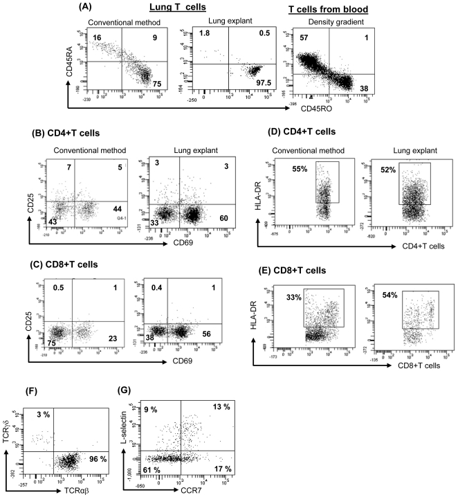
Recent studies have shown that tissue resident memory T cells (T(RM)) are critical to antiviral host defense in peripheral tissues. This new appreciation of T(RM) that reside in epithelial tissues and mediate host defense has been studied most extensively in skin: adult human skin contains large numbers of functional T(RM) that express skin specific markers. Indeed, more than twice as many T cells reside in skin as in peripheral blood. This T cell population has a diverse T cell receptor repertoire, and can produce a broad array of cytokines. More recently, we have begun to examine other epithelial tissues for the presence of resident T cells. In the present study, we asked whether analogous populations of resident T cells could be found in human lung. We were able to demonstrate abundant resident T cells in human lung-more than 10 billion T cells were present. Lung T cells were largely of the effector memory T cell (T(EM)) phenotype, though small numbers of central memory T cells (T(CM)) and T regulatory cells (T(reg)) could be identified. Lung T cells had a diverse T cell receptor repertoire and subsets produced IL-17, IL-4, IFNγ, as well as TNFα. A significant number of lung T(RM) CD4+Th cells produced more than one cytokine, identifying them as "multifunctional" Th1 type cells. Finally, lung T(RM), but not T(RM) resident to skin or T cells from blood, proliferated in response to influenza virus. This work suggests that normal human lung contains large numbers of T(RM) cells, and these cells are poised to respond to recall antigens previously encountered through lung mucosa. This population of T cells may contribute to the pathogenesis of asthma and other T cell mediated lung diseases.


Conflict of interest statement

Competing Interests: The authors have declared that no competing interests exist.

Figures

Figure 1
Figure 1. Human lung contains large numbers of T cells.
(A) Non-inflamed normal lung tissue was obtained and stained for hematoxylin and eosin (H&E), CD3 (200×), CD4 and CD8 (400×). A representative experiment is shown, and three additional donors produced similar results. (B) T cells were counted in sections 12 µm thick and 500 µm wide and estimated the numbers of T cells in 1 mm3. (C) After 3-day of explant culture, lung-T cells were harvested and counted as described in Materials & Methods. Data is shown as Mean ± SEM of 10 experiments. (D) Lung-T cells were extracted from same specimen by conventional as well as lung explant method and counted as described in Materials and Method section. Data is shown as Mean ± SEM of 3 experiments. (E) Lung-T cells, Blood T cells from freshly isolated PBMCs (negative control), or KG1a cells (positive control) were stained for Ki67 and analyzed by flow cytometry.
Figure 2
Figure 2. Direct comparison of conventional enzymatic methods and lung matrix explant methods.
T cells were extracted by conventional method and explant method. PBMCs were isolated by Ficoll-hypaque density gradient method. Cells were stained for surface markers and analyzed by flow cytometry. (A) Expression of CD45RO (TEM) and CD45RA on lung-CD3+T cells and blood-T cells was analyzed. (B–E) Many CD4 and CD8+T cells express CD69, HLA-DR and only few CD4+T cells but not CD8 express CD25. (F) Most of the lung-T cells are αβ+ and very few express γδ TCR. (G) A subpopulation of CD4+ CD45RA- T cells also express L-selectin and CCR7 (markers for central memory phenotype). Dot plots are representative of 9 experiments (3A–F) that produced similar results.
Figure 3
Figure 3. Lung T cells uniformly express VLA-1 and PSGL1 but not skin homing (CLA) and gut homing (α4β7) markers.
Lung T cells were extracted by lung explant method and stained for CLA (HECA-452) (A), α4β7 (ACT-1) (B), and PSGL-1 (C) and analyzed by flow cytometry. Lung-T cells, skin-T cells and gut-T cells were stained for VLA-1 (CD49a+CD29+) and analyzed by flow cytometry (D). A representative dot plot of each marker is shown and 3 experiments (α4β7, PSGL-1, VLA-1) and 2 experiments (CLA) produced similar results.
Figure 4
Figure 4. Large numbers of immunocompetent and influenza-specific T cells resides in human lung.
Lung T cells were extracted by lung explant method. (A–B) The cytokine secretion of effector memory T cells after overnight stimulation with artificial APCs (microbeads coated with anti-CD2, anti CD3 and antiCD28 mAbs) at 1∶1 cells: bead ratio. Brefeldin A (golgi-stop) was added 6 h prior to intracellular staining of cytokines. A representative dot plot of each cytokine is shown and 6 additional experiments produced similar results. (C) CD4+T cells were stained with TNFα, IL-2 and IFNγ after stimulation with PMA+ionomycin for 6h in presence of brefeldin A (Gate on CD4+IFNγ+ population). A representative dot plot is shown and 10 additional experiments produced similar results. (D) Lung T cells were isolated and stained for different Vbeta T cell receptors using TCR V beta repertoir kit (Beckman coulter) according to manufacturer's instructions. Diversity of V beta TCRs was analyzed by flow cytometry. Data represent Mean +/− SEM of 3 different donors. (E) CFSE labeled T cells from lung, skin and blood were cultured with heat killed influenza virus pulsed APCs in 1∶2 ratio. On day 4, T cell proliferation was measured by analyzing CFSE dilution using flow cytometry. A representative experiment is shown and 2 additional experiments produced similar results.

References

    1. Debes GF, Arnold CN, Young AJ, Krautwald S, Lipp M, et al. Chemokine receptor CCR7 required for T lymphocyte exit from peripheral tissues. Nat Immunol. 2005;6:889–894. - PMC - PubMed
    1. Luster AD, Alon R, von Andrian UH. Immune cell migration in inflammation: present and future therapeutic targets. Nat Immunol. 2005;6:1182–1190. - PubMed
    1. Robert C, Kupper TS. Inflammatory skin diseases, T cells, and immune surveillance. N Engl J Med. 1999;341:1817–1828. - PubMed
    1. Clark RA, Chong B, Mirchandani N, Brinster NK, Yamanaka K, et al. The vast majority of CLA+ T cells are resident in normal skin. J Immunol. 2006;176:4431–4439. - PubMed
    1. Clark RA, Chong BF, Mirchandani N, Yamanaka K, Murphy GF, et al. A novel method for the isolation of skin resident T cells from normal and diseased human skin. J Invest Dermatol. 2006;126:1059–1070. - PubMed

Publication types