

Predicted structures and dynamics for agonists and antagonists bound to serotonin 5-HT2B and 5-HT2C receptors
- PMID: 21299232
- PMCID: PMC3070210
- DOI: 10.1021/ci100375b
Predicted structures and dynamics for agonists and antagonists bound to serotonin 5-HT2B and 5-HT2C receptors
Abstract
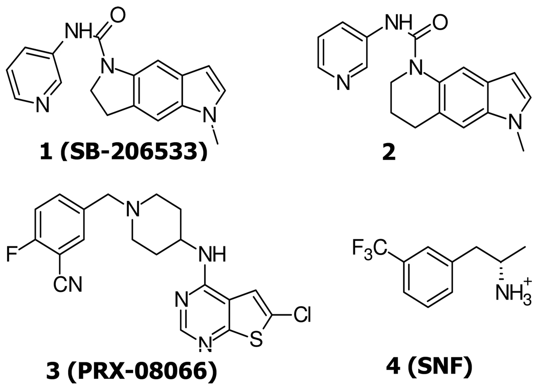
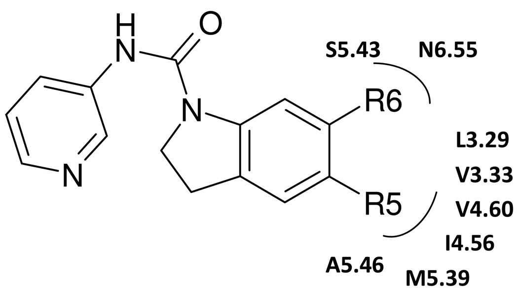
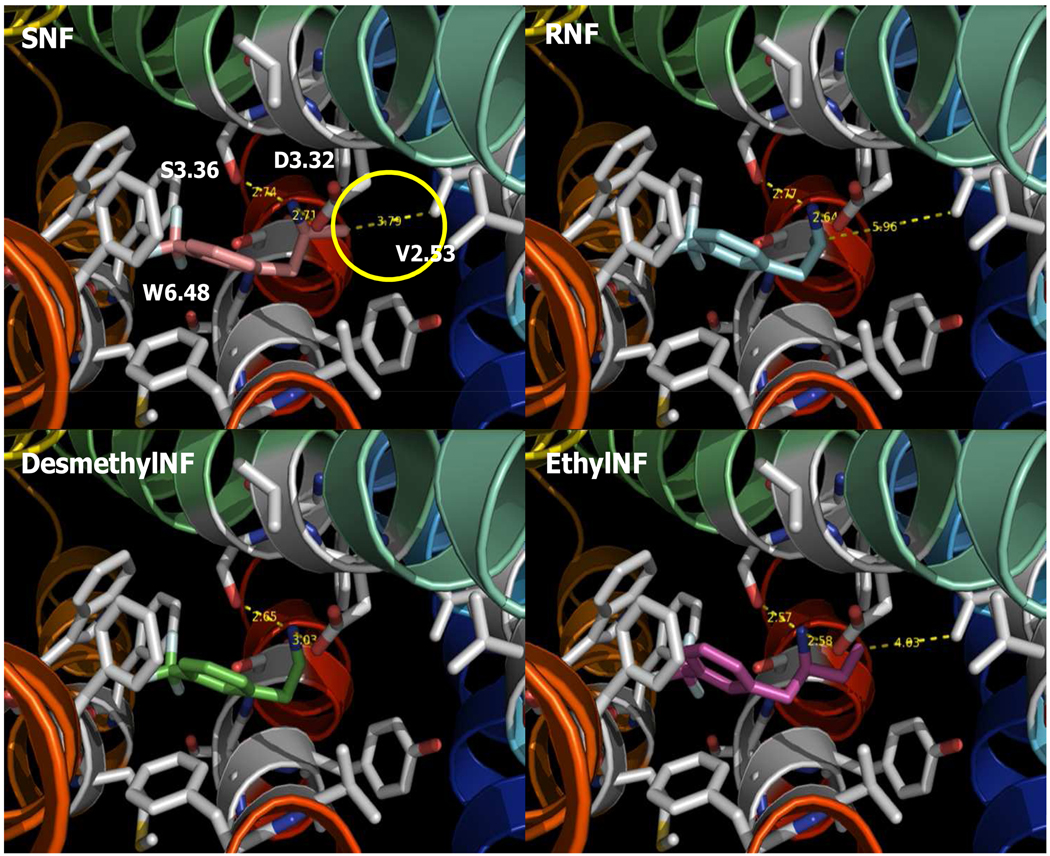
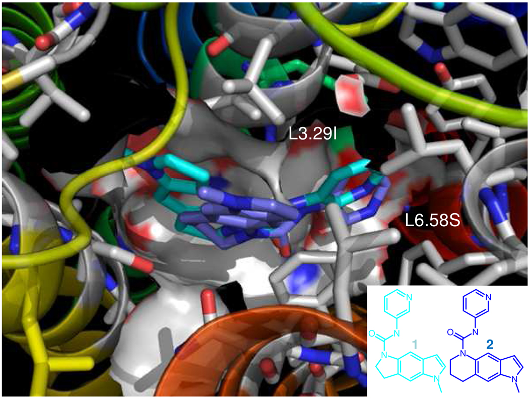
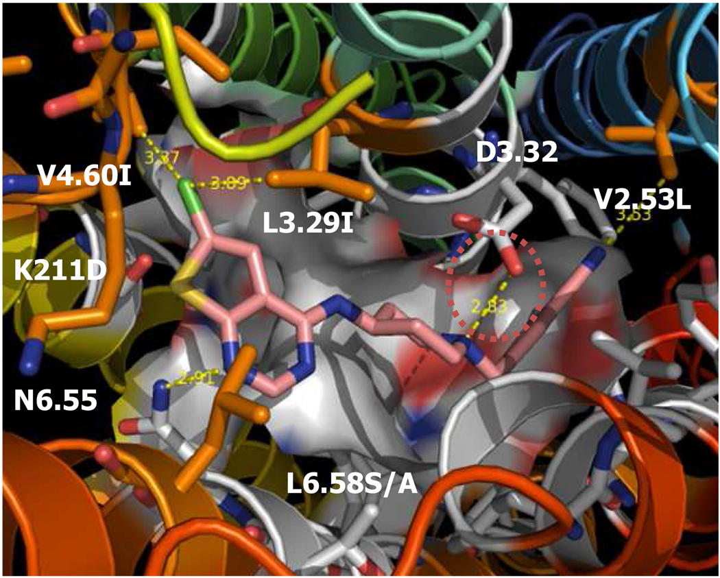
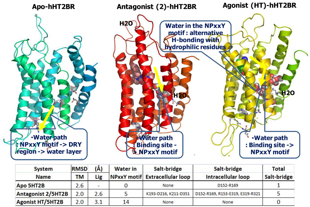
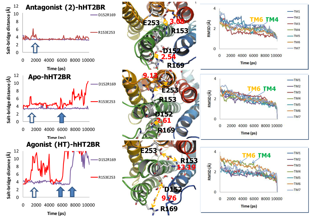
Subtype 2 serotonin (5-hydroxytryptamine, 5-HT) receptors are major drug targets for schizophrenia, feeding disorders, perception, depression, migraines, hypertension, anxiety, hallucinogens, and gastrointestinal dysfunctions. (1) We report here the predicted structure of 5-HT2B and 5-HT2C receptors bound to highly potent and selective 5-HT2B antagonist PRX-08066 3, (pKi: 30 nM), including the key binding residues [V103 (2.53), L132 (3.29), V190 (4.60), and L347 (6.58)] determining the selectivity of binding to 5-HT2B over 5-HT2A. We also report structures of the endogenous agonist (5-HT) and a HT2B selective antagonist 2 (1-methyl-1-1,6,7,8-tetrahydro-pyrrolo[2,3-g]quinoline-5-carboxylic acid pyridine-3-ylamide). We examine the dynamics for the agonist- and antagonist-bound HT2B receptors in explicit membrane and water finding dramatically different patterns of water migration into the NPxxY motif and the binding site that correlates with the stability of ionic locks in the D(E)RY region.
Figures
References
-
- Setola V, Dukat M, Glennon RA, Roth BL. Molecular determinants for the interaction of the Valvulopathic anorexigen norfenfluramine with the 5-HT2B receptor. Mol. Pharmacol. 2005;68:20–33. - PubMed
-
- Schmuck AG, Ullmer C, Engles P, Lübbert H. Cloning and Functional Characterization of The Human 5-HT2B Serotonin Receptor. FEBS Lett. 1994;342:85–90. - PubMed
-
- Bray JK, Goddard WA., III The structure of human serotonin 2c G-protein-coupled receptor bound to agonists and antagonists. J. Mol. Graph. Model. 2008;27:66–81. - PubMed
-
- Rasmussen SG, Choi HJ, Rosenbaum DM, Kobilka TS, Thian FS, Edwards PC, Burghammer M, Ratnala VR, Sanishvili R, Fischetti RF, Schertler GF, Weis WI, Kobilka BK. Crystal structure of the human beta2 adrenergic G-protein-coupled receptor. Nature. 2007;450:383–387. - PubMed
Publication types
MeSH terms
Substances
Grants and funding
LinkOut - more resources
Full Text Sources
Other Literature Sources
