. 2011 Feb;79(4):954-67.
doi: 10.1111/j.1365-2958.2010.07505.x. Epub 2010 Dec 22.

Function of the usher N-terminus in catalysing pilus assembly

Affiliations

Function of the usher N-terminus in catalysing pilus assembly

Nadine S Henderson et al. Mol Microbiol. 2011 Feb.

Abstract

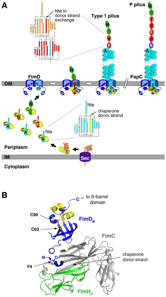
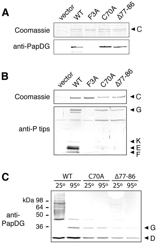
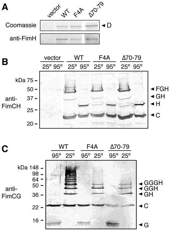
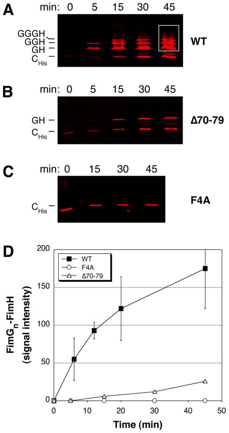
The chaperone/usher (CU) pathway is a conserved bacterial secretion system that assembles adhesive fibres termed pili or fimbriae. Pilus biogenesis by the CU pathway requires a periplasmic chaperone and an outer membrane (OM) assembly platform termed the usher. The usher catalyses formation of subunit-subunit interactions to promote polymerization of the pilus fibre and provides the channel for fibre secretion. The mechanism by which the usher catalyses pilus assembly is not known. Using the P and type 1 pilus systems of uropathogenic Escherichia coli, we show that a conserved N-terminal disulphide region of the PapC and FimD ushers, as well as residue F4 of FimD, are required for the catalytic activity of the ushers. PapC disulphide loop mutants were able to bind PapDG chaperone-subunit complexes, but did not assemble PapG into pilus fibres. FimD disulphide loop and F4 mutants were able to bind chaperone-subunit complexes and initiate assembly of pilus fibres, but were defective for extending the pilus fibres, as measured using in vivo co-purification and in vitro pilus polymerization assays. These results suggest that the catalytic activity of PapC is required to initiate pilus biogenesis, whereas the catalytic activity of FimD is required for extension of the pilus fibre.


Figures

Fig. 1
Fig. 1. Assembly of Type 1 and P pili by the CU pathway
A. Model for pilus assembly. The assembly steps for type 1 pili are shown, together with models for the fully assembled type 1 and P pili. The Fim and Pap proteins are indicated by single letters (H, FimH; etc.). Pilus subunits enter the periplasm as unfolded polypeptides via the Sec general secretory pathway. The subunits fold upon interaction with the periplasmic chaperone, forming stable complexes via donor strand complementation. Assembly and secretion of the pilus fiber occurs at the OM usher, where chaperone-subunit interactions are replaced with subunit-subunit interactions via the donor strand exchange reaction. During donor strand exchange, the Nte of an incoming chaperone-subunit complex displaces the chaperone donor strand from the preceding subunit. Topology diagrams are shown depicting the donor strand complementation and exchange reactions occurring with the chaperone and in the pilus fiber, respectively. The dimeric ushers are depicted with the central ß-barrel domain forming a channel that spans the OM, the N- and C-terminal domains (labeled N and C, respectively) located in the periplasm, and the plug domain (labeled P) gating the channel shut. Chaperone-adhesin complexes have highest affinity for the usher and initiate pilus assembly by binding to the usher N-terminal domain. The pilus tip fiber is assembled first, followed by the pilus rod. B. Structure of the FimDN-CHp complex (PDB ID: 1ZE3). FimC is shown in gray and FimHp in green. FimDN is shown in blue, with the disulfide loop region colored yellow. FimD residue F4 and the disulfide bond between residues C63 and C90 are shown in stick representation. The donated ß-strand of the FimC chaperone that occupies the FimHp subunit groove is indicated. The FimDN structure represents usher N-terminal domain cartooned in (A). The C-terminal end of the FimDN domain is connected by a linking region of 14 residues to the transmembrane ß-barrel domain of the usher. The structure was generated using PyMOL (http://www.pymol.org).
Fig. 2
Fig. 2. Analysis of the PapC N-terminal disulfide loop mutants
A. Overlay assay for binding of PapDG to PapC. OM fractions were isolated from strain SF100 expressing vector only, WT PapC, or the indicated PapC mutant. Duplicate samples were subjected to SDS-PAGE and either stained with Coomassie blue (upper panel) or transferred to a PVDF membrane for the overlay assay. Binding of PapDG to the usher (lower panel) was determined by immunoblotting with anti-PapDG antibodies. B. Co-purification of pilus tip subunits with PapC. OM fractions were isolated from strain SF100/pPAP58 (papDJKEFG) expressing vector only, WT PapC, or the indicated PapC mutant. His-tagged PapC was purified from the OM fractions, subjected to SDS-PAGE, and either stained with Coomassie blue to show the amount of PapC present (upper panel) or immunoblotted with anti-P pilus tips antiserum to detect pilus tip subunits that co-purified with the usher (PapG, K, E and F, lower panel). C. Assembly of PapG into pilus tip fibers. Co-purification of pilus tip subunits with WT PapC or the indicated PapC mutant was performed as in (B). The samples were incubated at 25 or 95°C in SDS-PAGE sample buffer, subjected to SDS-PAGE and immunoblotted with anti-PapDG antibodies.
Fig. 3
Fig. 3. Analysis of the FimD F4A and Δ70–79 mutants
A. Overlay assay for binding of FimCH to FimD. OM fractions were isolated from strain SF100 expressing vector only, WT FimD, or the indicated FimD mutant. Duplicate samples were subjected to SDS-PAGE and either stained with Coomassie blue (upper panel) or transferred to a PVDF membrane for the overlay assay. Binding of FimCH to the usher (lower panel) was determined by immunoblotting with anti-FimH antibody. B. Co-purification of FimCFGH pilus tip fibers with FimD. OM fractions were isolated from strain AAEC185/pNH235 (fimCFGH) expressing vector only, WT FimD, or the indicated FimD mutant. His-tagged FimD was purified from the OM fractions, incubated at 25 or 95°C in sample buffer, subjected to SDS-PAGE, and immunoblotted with anti-FimCH antibodies. The identities of the complexes that co-purified with FimD are indicated on the right using single letters to represent the Fim proteins (H, FimH; etc.). C. Co-purification of FimCGH pilus tip fibers with FimD. The assay was performed as in (B), except the host strain was AAEC185/pNH222 (fimCGH) and the samples were immunoblotted with anti-FimCG antibodies.
Fig. 4
Fig. 4. In vitro reconstitution assay for polymerization of type 1 pili
A–C. Reconstitution assay for pilus assembly. OM fractions were isolated from strain Tuner expressing WT FimD (A), FimD Δ70–79 (B), or FimD F4A (C). The OM fractions were mixed together with separately isolated periplasm fractions containing FimCH and FimCHisG and incubated at 25°C for the indicated time points. Pilus assembly intermediates purified from the OM fractions were incubated in sample buffer at 25°C, subjected to SDS-PAGE, and immunoblotted using anti-FimCG antibodies. The blots were analyzed using the Odyssey Infrared Imaging System. D. Quantitation of FimGn-FimH complex formation. The amount of FimGn-FimH complexes assembled at each time point in the reconstitution assay, preformed as in (A–C), was determined by measuring the signal intensity within identical areas boxed as shown in (A) for the 45 min time point. The values for WT FimD represent means ± standard deviation of at least 5 separate experiments; the values for the FimD Δ70–79 and F4A mutants represent means of 2 separate experiments.

Similar articles

Cited by

References

    1. Baga M, Norgren M, Normark S. Biogenesis of E. coli Pap pili: PapH, a minor pilin subunit involved in cell anchoring and length modulation. Cell. 1987;49:241–251. - PubMed
    1. Baneyx F, Georgiou G. In vivo degradation of secreted fusion proteins by the Escherichia coli outer membrane protease OmpT. J Bacteriol. 1990;172:491–494. - PMC - PubMed
    1. Barnhart MM, Sauer FG, Pinkner JS, Hultgren SJ. Chaperone-subunit-usher interactions required for donor strand exchange during bacterial pilus assembly. J Bacteriol. 2003;185:2723–2730. - PMC - PubMed
    1. Blomfield IC, McClain MS, Eisenstein BI. Type 1 fimbriae mutants of Escherichia coli K12: characterization of recognized afimbriate strains and construction of new fim deletion mutants. Mol Microbiol. 1991;5:1439–1445. - PubMed
    1. Choudhury D, Thompson A, Stojanoff V, Langermann S, Pinkner J, Hultgren SJ, Knight SD. X-ray structure of the FimC-FimH chaperone-adhesin complex from uropathogenic Escherichia coli. Science. 1999;285:1061–1066. - PubMed

Publication types

MeSH terms