. 2011 Mar;32(3):206-13.
doi: 10.1016/j.placenta.2011.01.006.

The effects of thrombin and cytokines upon the biomechanics and remodeling of isolated amnion membrane, in vitro

Affiliations

The effects of thrombin and cytokines upon the biomechanics and remodeling of isolated amnion membrane, in vitro

D Kumar et al. Placenta. 2011 Mar.

Abstract

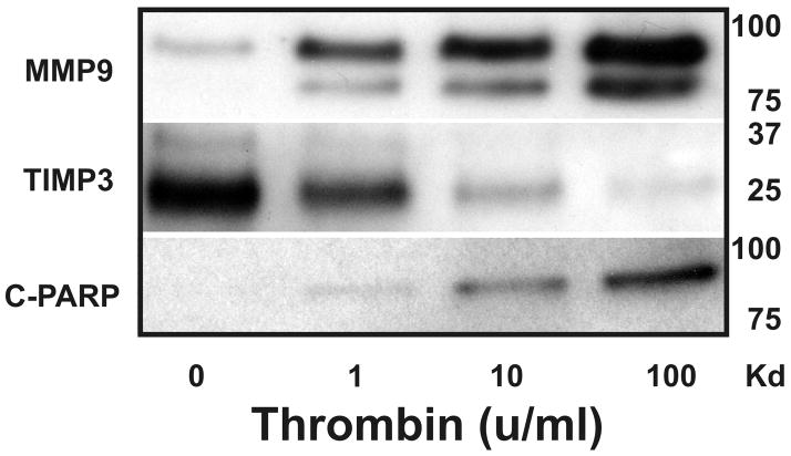
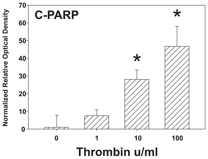
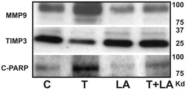
Abruption-induced thrombin generation and inflammation/infection induced cytokine production have both been associated with fetal membrane (FM) weakening and preterm premature rupture of the fetal membranes (PPROM). Using our in vitro model system we have demonstrated that thrombin, and separately the cytokines, tumor necrosis factor-alpha (TNFα) and interleukin-1-beta (IL-1β), remodel and weaken full thickness FM. Additionally, we have reported that the anti-oxidant and NFκB inhibitor, alpha-lipoic acid (LA), blocks these thrombin and cytokine induced effects. The purpose of these studies was to determine whether thrombin and cytokines directly weaken the amnion membrane (AM), the major load-bearing component of FM. Isolated AM or full thickness FM fragments from unlabored Cesarean deliveries were incubated with thrombin, TNFα, or IL-1β, for 48 h. Rupture strength (breaking force) of each fragment was thereafter determined using our published methodology. Biochemical evidence of remodeling and apoptosis; immunoreactive Matrix Metalloproteinase 9 (MMP9), Tissue Inhibitor of Matrix Metalloproteinase 3 (TIMP3) and cleaved poly (ADP-ribose) polymerase (C-PARP) levels in tissue extracts, were determined by western blot and densitometry. Thrombin induced a dose-dependent weakening of isolated AM (P < 0.001) coupled with dose dependent increases in PARP cleavage, and reciprocal increases and decreases, respectively, in MMP9 and TIMP3 protein (all P < 0.01). Thrombin receptor activating peptide-6 (TRAP) also weakened isolated AM. Neither TNFα nor IL-1β weakened isolated AM. However, both cytokines weakened AM when it was incubated together with the choriodecidua as part of full thickness FM (P < 0.001). Cytokine-conditioned choriodecidua medium also weakened isolated AM (P < 0.001). Under conditions in which cytokines weakened the AM, the changes in MMP9, TIMP3 and PARP cleavage were consistent with those seen after thrombin incubation. LA blocked the FM weakening and remodeling effects. In summary, thrombin weakens AM directly whereas cytokines weaken AM indirectly by causing the release of soluble intermediates from the choriodecidua.


Figures

Figure 1
Figure 1. Effect of Increasing Thrombin Dose upon AM Mechanical Properties
Thrombin (0–100 u/ml) induced concentration-dependent decreases in FM rupture strength. All incubations were for 48 h. Each data point shown represents the mean ± SD for four experiments (using different patient FM) with three replicates/experiment (N=12; *P < 0.001).
Figure 2
Figure 2. The Thrombin receptor (PAR1) activating peptide TRAP weakens Isolated AM
TRAP (1.0 μM) induces significant weakening of cultured AM following 48 h incubation. Each data point shown represents the mean ± SD for three experiments (using different patient FM) with three replicates/experiment (N=9; *P < 0.001).
Figure 3
Figure 3. Effect of Increasing Thrombin Dose upon AM Remodeling and Apoptosis
Thrombin (0–100 u/ml for 48 h) induced increases in both pro-MMP9 (92 kD) and active MMP9 (78 kD) proteins (upper panel) and decreased TIMP3 (27 kD and 24 kD) proteins (middle panel) in isolated AM fragments in a dose-dependent manner. Thrombin (0–100 u/ml for 48 h) induced apoptosis in AM explants as evidenced by cleavage of PARP (113 kD) into 85 kD (and 24 kD; not shown) (lower panel) fragments. A: Representative western blot. B: To confirm the significance of qualitative results, blots were scanned and subjected to densitometric analysis. Each blot was normalized to the zero-thrombin controls as indicated in Methods. Data points represent normalized data from four experiments each with three replicates; mean ± SD; N=12, (* P < 0.01).
Figure 4
Figure 4. Cytokines Mediate AM Weakening Through Choriodecidua
A. Study Design: AM strength, MMP9 protein, TIMP3 protein and PARP cleavage were measured after incubation with control medium, 50 ng/ml TNFα, or 50 ng/ml IL-1β under one of three conditions: 1. Full thickness FM were incubated for 72 h followed by separation and testing of the AM; 2. Isolated AM was incubated for 72 h and then tested; 3. Isolated choriodecidua was incubated for 24 h after which the medium was transferred to isolated AM in culture for an additional 72 h and then tested. B. Results: Upper panel: TNFα or IL-1β decreased AM rupture strength if the AM was incubated together with choriodecidua directly in intact, full thickness FM (left) or through cytokine-conditioned medium from choriodecidua (right), but not if isolated AM was incubated with the cytokines (data are presented as mean ± SD; N=12, * P < 0.001). Lower panel: Representative western blots for MMP9, TIMP3 and Cleaved PARP are shown for the same conditions as Rupture Strength data in the upper panel. MMP9 and PARP cleavage increase and TIMP3 decreases in parallel with weakening.
Figure 5
Figure 5. LA inhibits Thrombin-induced Weakening
AM fragments were pre-incubated with or without LA (0.25 mM) for 6h, then incubated with or without the addition of Thrombin (10 u/ml) for 48h. Four groups are displayed: Control, Thrombin alone, LA pre-treatment alone, LA pretreatment + Thrombin. Rupture Strength was determined as outlined in Methods. Data points represent pooled results of three experiments using three FM fragments per treatment group in each experiment (N=9). Data are presented as mean ± SD (* P <0.001).
Figure 6
Figure 6. LA inhibits Thrombin induced remodeling in Isolated AM
A. Isolated AM fragments were pre-incubated with or without LA (0.25 mM) for 6h, then incubated with or without the addition of thrombin (10 u/ml) for 48h. Four groups are displayed: Control, Thrombin alone, LA pre-treatment alone, LA pretreatment + Thrombin. The Western blot shown is representative of three replicate experiments with three different placentas (N=9). Both the pro-MMP9 (92 kD) and the active MMP9 (78 kD) protein, as well as TIMP3 (27 kD and 24 kD) and cleaved PARP are shown. B. To confirm the significance of qualitative results, blots were scanned and subjected to densitometric analysis. Each blot was normalized to the zero-thrombin controls as indicated in Methods (Data are presented mean ± SD; N=9, * P < 0.05).

References

    1. Mercer BM. Premature Rupture of the membranes. An expert’s view. Obstet G ynecol. 2003;101:178–93. - PubMed
    1. Menon R. Spontaneous preterm birth, a clinical dilemma: etiologic, pathophysiologic and genetic heterogeneities and racial disparity. Acta Obstet Gynecol Scand. 2008;87:590–600. - PubMed
    1. Parry S, Strauss JF. Premature rupture of the fetal membranes. N Engl J Med. 1998;338:663–70. - PubMed
    1. Menon R, Fortunato SJ. The role of matrix degrading enzymes and apoptosis in rupture of membranes. J Soc Gynecol Investig. 2004;11:427–37. - PubMed
    1. Moore RM, Mansour JM, Redline RW, Mercer BM, Moore JJ. The physiology of fetal membrane rupture: insight gained from the determination of physical properties. Placenta. 2006;27:1037–51. - PubMed

Publication types

MeSH terms