. 2011 Apr 1;589(Pt 7):1663-80.
doi: 10.1113/jphysiol.2010.201491. Epub 2011 Feb 7.

Is connexin36 critical for GABAergic hypersynchronization in the hippocampus?

Affiliations

Is connexin36 critical for GABAergic hypersynchronization in the hippocampus?

Michael Beaumont et al. J Physiol. .

Abstract

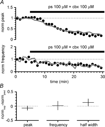
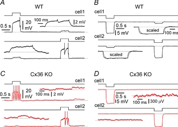
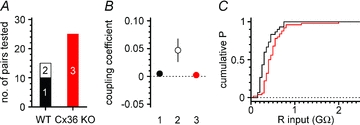
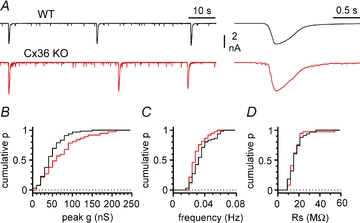
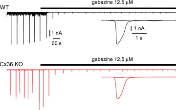
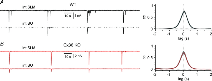
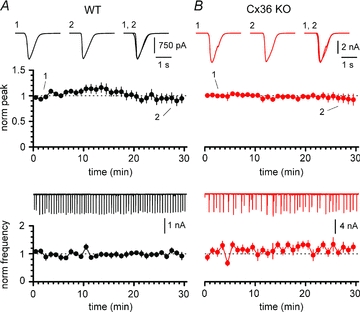
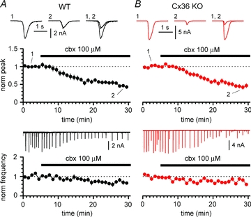
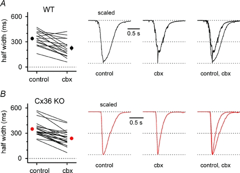
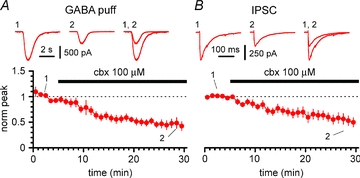
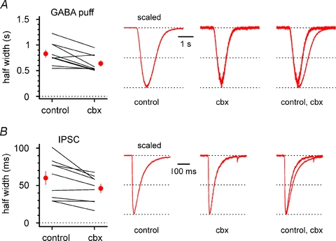
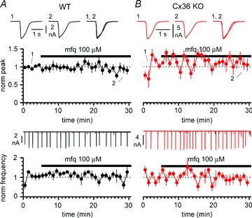
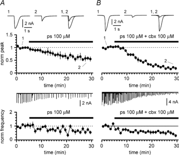
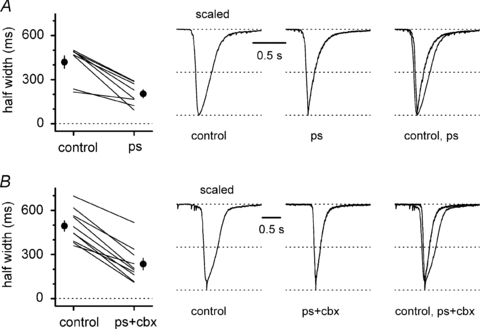
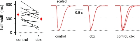
Synchronous bursting of cortical GABAergic interneurons is important in epilepsies associated with excitatory GABAergic signalling. If electrical coupling was critical for the generation of this pathological activity, then the development of selective blockers of connexin36-based interneuronal gap junctions could be of therapeutic value. We have addressed this issue in the 4-aminopyridine model of epilepsy in vitro by comparing GABAergic epileptiform currents and their sensitivity to gap junction blockers in wild-type vs. connexin36 knockout mice. Although electrical coupling was abolished in stratum lacunosum-moleculare interneurons from knockout animals, epileptiform currents were not eliminated. Furthermore, epileptiform currents propagated similarly across hippocampal layers in the two genotypic groups. Blockade of electrical coupling with carbenoxolone suppressed amplitude, frequency and half-width of the epileptiform currents both in wild-type and in knockout animals, whereas mefloquine had no effects. Carbenoxolone also depressed responses to exogenous and synaptic GABA application onto interneurons. We conclude that, in the 4-aminopyridine model of epilepsy in vitro, connexin36 is not critical for the generation of epileptiform discharges in GABAergic networks and that the observed antiepileptic effects of carbenoxolone are likely to be due to blockade of GABAA receptors and not of connexin36-based gap junctions. Lastly, because of its chemical structure and its effects on amplitude and kinetics of GABAergic currents, we tested the hypothesis that carbenoxolone acted via specific sites on GABAA receptors, such as the one mediating the effects of the neurosteroid pregnenolone sulfate, or the allosteric regulatory site of benzodiazepines/β-carbolines. Our results suggest that neither of these is involved.


Figures

Figure 14
Figure 14. The effects of pregnenolone sulfate and carbenoxolone on 4-AP-induced epileptiform events add linearly
A, time course plot of the experimental effect of co-application of pregnenolone sulfate and carbenoxolone on amplitude and frequency of GABAergic currents (black circles, same data as Fig. 12B) compared to its predicted effect calculated from the linear summation of the effects of their individual applications (thick grey line). Notice the similarity of the effects. B, mean and 95% confidence intervals of the difference of the experimental effect of co-application (from left to right: on peak amplitude, event frequency and half-width) vs. the predicted effect if the two drugs act by purely independent mechanisms. Notice that all the confidence intervals of the difference (normexp– normpred) include a range of values indicating a very small difference between observed and predicted effects.
Figure 1
Figure 1. Electrical coupling between CA1 stratum lacunosum-moleculare interneurons in wild-type (WT, black traces) and connexin36 knockout (Cx36 KO, red traces) mice
A, sequential injection of a depolarizing current step (90 pA, black steps) to interneurons of WT mice generates an attenuated response in the non-injected cell. Insets show the attenuated responses at enhanced magnification: notice the presence of spikelets. B, similar protocol as in A, but hyperpolarizing current steps of –50 pA (black steps) are injected. Notice the propagation of the voltage response to the non-injected neuron. Also notice, in the insets, after scaling, the slower kinetics of the propagated response (dotted line) vs. the original one (continuous trace). C and D illustrate the same type of experiments as A and B, respectively, performed on interneurons in a slice obtained from a Cx36 KO mouse. Notice the lack of a propagated response in the non-injected neuron. Current pulses were 50 pA in C (black steps) and –50 pA in D (black steps). Insets in C and D show the lack of a voltage response in the non-injected cell at a magnified scale. Gabazine (12.5 μm), NBQX (20 μm), d-AP5 (50 μm) and CGP55845 (1–5 μm) present throughout in AD.
Figure 2
Figure 2. Network and membrane differences in interneurons of wild-type (WT) vs. connexin36 knockout (Cx36 KO) mouse
A, summary plot showing the different probability of finding electrically coupled pairs in the two types of animals. The white part of the bar (2) represents coupled pairs, in contrast to the black and red portions (1, 3), representing non-connected neurons in wild-type and Cx36 KO mice, respectively. B, summary graph showing the value of coupling coefficients for the uncoupled (1, black circle) and coupled (2, white circle) pairs of WT animals and for uncoupled (3, red circle) cells of Cx36 KO mice. C, cumulative probability distributions of resting membrane input resistances in WT (black line) vs. Cx36 KO animals (red line). Notice the shift towards higher values in the Cx36 KO mice.
Figure 3
Figure 3. Properties of 4-AP-induced epileptiform currents and recording conditions in WT (black traces) vs. Cx36 KO mice (red traces)
A, examples of voltage-clamp recording traces from stratum lacunosum-moleculare interneurons in WT and Cx36 KO mice shown at different temporal magnifications (left and right panels). B, cumulative probability plot of the peak conductance during epileptiform events in the two genotypes (black line, WT; red line, Cx36 KO). Notice the presence of larger conductance events in the Cx36 KO genotype. C, summary graph showing the similarity of the frequency of the epileptiform events in the two groups of animals. D, cumulative probability plot of the series resistance for the recordings in WT and Cx36 KO mice: notice the overlapping distributions. AD, ionotropic glutamatergic synaptic transmission and GABAB receptors blocked throughout the experiments.
Figure 4
Figure 4. Pharmacological demonstration that 4-AP-induced epileptiform currents are mediated by GABAA receptors in both WT (black traces) and Cx36 KO mice (red traces)
Application of the GABAA receptor blocker gabazine (12.5 μm) powerfully reduces epileptiform currents in interneurons recorded in slices obtained from the two different genotypes. Upper traces show wild-type and lower traces connexin36 knockout. Insets show superimposed averaged events in control and in the presence of the drug.
Figure 5
Figure 5. Propagation of 4-AP-induced epileptiform currents across hippocampal layers in WT (black traces) vs. Cx36 KO animals (red traces)
A, left panel, simultaneous recordings from a stratum lacunosum-moleculare and a stratum oriens interneuron from a WT mouse (int SLM and int SO, respectively). Notice the presence of synchronous events. Right panel, summary cross-correlogram of the activity in 10 SLM-SO WT interneuron pairs. Grey bands indicate ±SEM. B, identical experiments and analysis as in A, but performed in 10 pairs of interneurons from Cx36 KO animals. Notice the similarity of the results. NBQX (20 μm), d-AP5 (50 μm) and CGP55845 (5 μm) present throughout in A and B.
Figure 6
Figure 6. 4-AP-induced epileptiform activity can be maintained for long recording times in both WT (black circles and traces) and Cx36 KO mice (red circles and traces)
A, notice the stability of the amplitude (upper panel) and frequency (lower panel) of the events. Insets in the upper panel represent averaged events during the first (1) and last (2) 5 min of recording, and superimposed (1, 2). Inset in the lower panel shows the continuous experimental recording. B, as in A, but experiments were performed in slices from Cx36 KO animals. Note that epileptiform activity is similarly maintained for 30 min in both genotypes. NBQX (20 μm), d-AP5 (50 μm) and CGP55845 (5 μm) present throughout in A and B.
Figure 7
Figure 7. Carbenoxolone depresses GABAergic epileptiform events in both WT (black circles and traces) and Cx36 KO animals (red circles and traces)
A, time course of normalized peak (upper panel) and frequency (lower panel) during application of carbenoxolone (cbx, black bar) to slices from WT mice. Notice the strong decrease in the amplitude of the events and also the reduction in their frequency. Insets in the upper panel are averaged events in control (1), in the presence of carbenoxolone (2), and superimposed (1, 2). Inset in the lower panel shows the continuous experimental recording. B, as in A, but experiments were performed on Cx36 KO animals. Notice that carbenoxolone also has an effect on both amplitude and frequency in this genotype. Ionotropic glutamatergic synaptic transmission and GABAB receptors blocked throughout the experiments both in A and B.
Figure 8
Figure 8. Carbenoxolone reduces the half-width of GABAergic epileptiform events in both WT (black circles and traces) and Cx36 KO animals (red circles and traces)
A, left, summary graph comparing half-widths in control and in the presence of carbenoxolone (cbx) for individual experiments (lines) and for their overall average (circle). Traces in control, in the presence of carbenoxolone, and superimposed are shown in the right panel. B, as in A, but experiments were performed on slices from Cx36 KO animals. Notice that carbenoxolone has a similar effect irrespective of the genotype. NBQX (20 μm), d-AP5 (50 μm) and CGP55845 (5 μm) present throughout in A and B.
Figure 9
Figure 9. Carbenoxolone reduces direct responses both to exogenous and synaptic GABA applications
A, summary plot of the time course of the effect of carbenoxolone (cbx, black bar) on currents evoked by GABA puffs onto stratum lacunosum-moleculare interneurons of the Cx36 KO mouse. Insets show averaged responses to GABA application in control (1), in the presence of carbenoxolone (2) and superimposed (1, 2). B, time course summary graph for the effect of carbenoxolone (black bar) on evoked inhibitory postsynaptic currents (IPSCs) recorded from the same cells in A. Notice that IPSCs are also depressed by carbenoxolone, similarly to responses to exogenous GABA. All experiments in A and B were performed on slices from Cx36 KO animals and ionotropic glutamatergic synaptic transmission and GABAB receptors were pharmacologically blocked.
Figure 10
Figure 10. Effect of carbenoxolone on the half-width of responses to exogenous GABAergic applications and of inhibitory postsynaptic potentials in interneurons from Cx36 KO animals
A, left, summary graph comparing half-widths of voltage-clamp responses to GABA puffs in control and in the presence of carbenoxolone (cbx) for individual experiments (lines) and for their overall average (red circles). Traces in control, in the presence of carbenoxolone and superimposed are shown in the right panel. B, as in A, but data and analysis regards evoked inhibitory postsynaptic currents (IPSCs). Notice that carbenoxolone has a similar effect on GABA puffs and IPSCs. NBQX (20 μm), d-AP5 (50 μm) and CGP55845 (5 μm) present throughout in A and B.
Figure 11
Figure 11. Mefloquine does not affect GABAergic epileptiform events in either WT (black circles and traces) or Cx36 KO animals (red circles and traces)
A, time course of normalized peak (upper panel) and frequency (lower panel) during application of mefloquine (mfq, black bar) to slices from WT mice. Notice the lack of effect both on the amplitude and on the frequency of the events. Insets in the upper panel are averaged events in control (1), in the presence of mefloquine (2), and superimposed (1, 2). Inset in the lower panel shows the continuous experimental recording. B, as in A, but experiments were performed on Cx36 KO animals. Notice that mefloquine does not affect epileptiform activity in this genotype either. Ionotropic glutamatergic synaptic transmission and GABAB receptors blocked throughout the experiments in both A and B.
Figure 12
Figure 12. Effects of the application of pregnenolone sulfate on 4-AP-induced epileptiform currents and of its co-application with carbenoxolone in slices from WT animals
A, time course of normalized peak (upper panel) and frequency (lower panel) during application of pregnenolone sulfate (ps, black bar). Notice the reduction in both amplitude and frequency of the events. Insets in the upper panel are averaged events in control (1), in the presence of pregnenolone sulfate (2), and superimposed (1, 2). Inset in the lower panel shows the continuous experimental recording. B, as in A, but pregnenolone sulfate and carbenoxolone (ps+cbx) were co-applied. Ionotropic glutamatergic synaptic transmission and GABAB receptors blocked throughout the experiments both in A and B.
Figure 13
Figure 13. Kinetic changes of 4-AP-induced epileptiform currents following either application of pregnenolone sulfate or its co-application with carbenoxolone in slices from WT animals
A, left, summary graph comparing half-widths in control and in the presence of pregnenolone sulfate (ps) for individual experiments (lines) and for their overall average (black circles). Traces in control, in the presence of pregnenolone sulfate and superimposed are shown in the right panel. B, as in A, but pregnenolone sulfate and carbenoxolone were co-applied. Ionotropic glutamatergic synaptic transmission and GABAB receptors were pharmacologically blocked in A and B.
Figure 15
Figure 15. Carbenoxolone does not act via the allosteric regulatory site of benzodiazepines
A, time course of normalized peak (upper panel) and frequency (lower panel) during the application of carbenoxolone (cbx, black bar) in the constant presence of the competitive antagonist flumazenil: notice the similarity of these results (red circles) to those obtained in the absence of flumazenil (thick grey lines, same data as in Fig. 7B). Insets in the upper panel are averaged events in flumazenil (1), in the presence of flumazenil and carbenoxolone (2), and superimposed (1, 2). Inset in the lower panel shows the continuous experimental recording. B, carbenoxolone does not change GABAergic epileptiform currents at low concentrations. Notice the lack of effects on both amplitude and frequency. Insets in the upper panel are averaged events in control (1), in the presence of low concentrations of carbenoxolone (2), and superimposed (1, 2). Inset in the lower panel shows the continuous experimental recording. Experiments shown both in A and B were performed in slices obtained from Cx36 KO mice and in the constant additional presence of NBQX (20 μm), d-AP5 (50 μm) and CGP55845 (5 μm).
Figure 16
Figure 16. Kinetic changes of 4-AP-induced epileptiform currents following application of carbenoxolone in the constant presence of flumazenil
Left, summary graph comparing half-widths in flumazenil (5 μm, control) and in the added presence of carbenoxolone (100 μm, cbx) for individual experiments (lines) and for their overall average (red circles). Scaled traces in flumazenil, in the presence of flumazenil and carbenoxolone, and superimposed are shown in the right panel. Notice that flumazenil does not prevent carbenoxolone-induced kinetic changes.

References

    1. Avoli M. GABA-mediated synchronous potentials and seizure generation. Epilepsia. 1996;37:1035–1042. - PubMed
    1. Avoli M, Barbarosie M, Lücke A, Nagao T, Lopantsev V, Köhling R. Synchronous GABA-mediated potentials and epileptiform discharges in the rat limbic system in vitro. J Neurosci. 1996;16:3912–3924. - PMC - PubMed
    1. Avoli M, D'Antuono, Louvel J, Kohling R, Biagini G, Pumain R, D'Arcangelo G, Tancredi V. Network and pharmacological mechanisms leading to epileptiform synchronization in the limbic system in vitro. Prog Neurobiol. 2002;68:167–207. - PubMed
    1. Belelli D, Lambert JJ. Neurosteroids: endogenous regulators of the GABAA receptor. Nat Rev Neurosci. 2005;6:565–575. - PubMed
    1. Bennett MVL. Electrical transmission: a functional analysis and comparison to chemical transmission. In: Kandel EK, editor. Handbook of Physiology, section 1, The Nervous System, vol. I, Cellular Biology of Neurons. Baltimore: Williams & Wilkins; 1977. pp. 357–416.

Publication types

MeSH terms

LinkOut - more resources