. 2011 Feb 22;108(8):3330-5.
doi: 10.1073/pnas.1010890108. Epub 2011 Feb 7.

Skint-1 is a highly specific, unique selecting component for epidermal T cells

Affiliations

Skint-1 is a highly specific, unique selecting component for epidermal T cells

Susannah D Barbee et al. Proc Natl Acad Sci U S A. .

Abstract

αβ T-cell repertoire selection is mediated by peptide-MHC complexes presented by thymic epithelial or myeloid cells, and by lipid-CD1 complexes expressed by thymocytes. γδ T-cell repertoire selection, by contrast, is largely unresolved. Mice mutant for Skint-1, a unique Ig superfamily gene, do not develop canonical Vγ5Vδ1(+) dendritic epidermal T cells. This study shows that transgenic Skint-1, across a broad range of expression levels, precisely and selectively determines the Vγ5Vδ1(+) dendritic epidermal T-cell compartment. Skint-1 is expressed by medullary thymic epithelial cells, and unlike lipid-CD1 complexes, must be expressed by stromal cells to function efficiently. Its unusual transmembrane-cytoplasmic regions severely limit cell surface expression, yet increasing this or, conversely, retaining Skint1 intracellularly markedly compromises function. Each Skint1 domain appears nonredundant, including a unique decamer specifying IgV-domain processing. This investigation of Skint-1 biology points to complex events underpinning the positive selection of an intraepithelial γδ repertoire.


Conflict of interest statement

The authors declare no conflict of interest.

Figures

Fig. 1.
Fig. 1.
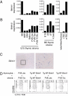
Ubiquitous Tg.NF-Skint1 expression restores Vγ5Vδ1+ DETC development. (A) Flow cytometry of thymocyte suspensions prepared from FVB.Jax, FVB.Tac, and FVB.Tac Tg.NF-Skint1−/+ line 39 fetuses at E17. Data are gated to show only Vγ5Vδ1+ (17D1+) thymocytes. Similar results were observed in all Tg.NF-Skint1 founder lines. (B) Flow cytometry of epidermal suspensions prepared from body wall skin of 3-wk-old mice. The bottom panels show γδTCR+ cells only. (C) Staining of DETC in FVB.Jax and Tg.NF-Skint1 epidermal sheets. (Scale bar: 20 μm.) (D and E) Quantitative RT-PCR analysis of Skint-1 expression in pooled E17 thymi and in skin from individual animals from Tg.NF-Skint1 (D) and untagged Skint1 (E) Tg lines, calculated relative to GAPDH. (F and G) Flow cytometry analysis of Skint1 Tg animals versus B6xSJL NLCs, performed as in A and B.
Fig. 2.
Fig. 2.
Tg NF-Skint1 does not regulate γδTCR repertoires outside the skin. (A) Western blot of Tg.NF-Skint1 protein in tissues (T, thymus; S, spleen; G, gut) from founder line 39. Protein was immunoprecipitated from Tg.NF-Skint1 and Tg.NF-Skint1+ tissue lysates with mouse anti-FLAG M2 antibody, and immunoblotted with rabbit anti-FLAG antibody. Putative monomer (◀) and dimer species (◁) are observed in all Tg tissues. (BE) Flow cytometry of cell suspensions prepared from the thymus, spleen, gut, and uterus of Tg and Tg+ animals of Tg.NF-Skint1 line 39. In B and C, all cells shown are gated on γδTCR+ and numbers reflect the percentage of γδ T cells, whereas CD3+ cell and total live cells are shown in D and E, respectively. (D) 17D1 binds to γδTCR+ cells that are Vγ5; this corresponds to a demonstrated GL3-dependent cross-reactivity with the Vγ6Vδ1+ TCR used by the uterine IEL compartment (33).
Fig. 3.
Fig. 3.
Skint-1 is expressed in thymic medullary epithelium in a TCR-independent fashion and effects function from a stromal cell context. (A) Quantitative RT-PCR analysis of Skint-1 expression in CD45 thymic stroma (Left and Center) or FACS-purified TEC (Right). CD45EpCam+ TEC from pooled FVB.Jax E15-E18 thymi were divided into Ly51+ cTEC and Ly51 mTEC subsets, and compared with total E17 thymus. Skint-1 expression is relative to GAPDH. (B) Quantitative RT-PCR analysis of Skint-2 expression as in A. (C) In situ hybridization analysis of Skint-1 expression in adult FVB.Jax thymus. Right: Close-up of medullary area indicated (Left). (D) Analysis of Vγ5Vδ1+ DETC precursor maturation (17D1+CD45RBhi) in mixed RTOC of purified CD45+ thymocyte progenitors and CD45 stroma from FVB.Jax, FVB.Tac, or Tg.NF-Skint1 E15 thymi.
Fig. 4.
Fig. 4.
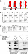
Biological and biochemical properties of Skint1. (A) Graphic representation of the Skint-1 constructs tested. (B) Surface flow cytometry of 293 cells transfected with the constructs in bicistronic vectors. All constructs include a FLAG tag at the N terminus and the KYVERTELL motif in the IgV domain; hCD2 is the marker of transduction efficiency. (C) Analysis of Vγ5Vδ1+ DETC precursor maturation in FVB.Tac RTOC 12 d after transduction with the constructs from A. (D) The multimerization of WT and Tac alleles is analyzed by Western blot analysis of 293 transfectants. Protein was immunoprecipitated with mouse anti-FLAG M2 or mouse anti-HA antibody, and immunoblotted with rabbit anti-FLAG antibody.
Fig. 5.
Fig. 5.
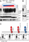
Proteolytic processing specified by IgC is required for function. (A and B) Western blot analysis of 293 cells transfected with N-terminal FLAG-tagged Skint-1/Skint-2 chimeric constructs. Protein was immunoprecipitated with mouse anti-FLAG M2 and immunoblotted with rabbit anti-FLAG antibody. In A, the recombination between Skint1 and Skint2 was shifted in 10-aa increments in the IgC, whereas in B, only the first 10 aa of the Skint1 IgC was replaced with the analogous sequence from Skint2. (C) Analysis of Vγ5Vδ1+ DETC precursor maturation in FVB.Tac RTOC 12 d after transduction with the indicated chimeric constructs.

References

    1. Hayday AC, Theodoridis E, Ramsburg E, Shires J. Intraepithelial lymphocytes: Exploring the Third Way in immunology. Nat Immunol. 2001;2:997–1003. - PubMed
    1. Havran WL, Allison JP. Developmentally ordered appearance of thymocytes expressing different T-cell antigen receptors. Nature. 1988;335:443–445. - PubMed
    1. Hayday A, Tigelaar R. Immunoregulation in the tissues by gammadelta T cells. Nat Rev Immunol. 2003;3:233–242. - PubMed
    1. Swamy M, Jamora C, Havran W, Hayday A. Epithelial decision makers: In search of the ‘epimmunome’. Nat Immunol. 2010;11:656–665. - PMC - PubMed
    1. Hayday AC. Gammadelta T cells and the lymphoid stress-surveillance response. Immunity. 2009;31:184–196. - PubMed

Publication types

Substances