. 2011 Feb 22;108(8):3395-400.
doi: 10.1073/pnas.1014411108. Epub 2011 Feb 7.

Proper formation of whisker barrelettes requires periphery-derived Smad4-dependent TGF-beta signaling

Affiliations

Proper formation of whisker barrelettes requires periphery-derived Smad4-dependent TGF-beta signaling

Susana da Silva et al. Proc Natl Acad Sci U S A. .

Abstract

Mammalian somatosensory topographic maps contain specialized neuronal structures that precisely recapitulate the spatial pattern of peripheral sensory organs. In the mouse, whiskers are orderly mapped onto several brainstem nuclei as a set of modular structures termed barrelettes. Using a dual-color iontophoretic labeling strategy, we found that the precise topography of barrelettes is not a result of ordered positions of sensory neurons within the ganglion. We next explored another possibility that formation of the whisker map is influenced by periphery-derived mechanisms. During the period of peripheral sensory innervation, several TGF-β ligands are exclusively expressed in whisker follicles in a dynamic spatiotemporal pattern. Disrupting TGF-β signaling, specifically in sensory neurons by conditional deletion of Smad4 at the late embryonic stage, results in the formation of abnormal barrelettes in the principalis and interpolaris brainstem nuclei and a complete absence of barrelettes in the caudalis nucleus. We further show that this phenotype is not derived from defective peripheral innervation or central axon outgrowth but is attributable to the misprojection and deficient segregation of trigeminal axonal collaterals into proper barrelettes. Furthermore, Smad4-deficient neurons develop simpler terminal arbors and form fewer synapses. Together, our findings substantiate the involvement of whisker-derived TGF-β/Smad4 signaling in the formation of the whisker somatotopic maps.


Conflict of interest statement

The authors declare no conflict of interest.

Figures
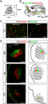
Fig. 1.
Fig. 1.
Trigeminal sensory neurons innervating neighboring whiskers are intermingled and scattered in the TG. (A) Schematic diagram of the topographic arrangement of large whiskers in the mouse face. Whiskers are organized in five rows (A–E) along the D-V axis and in up to seven arcs (1–7) along the A-P axis. (B) D2 and D3 whiskers were injected with Dextran-fluorophore tagged with either Alexa568 or Alexa488 at P0. Injections resulted in labeling of cell bodies and axon projections of trigeminal sensory neurons. (C) Horizontal and sagittal sections of TG 3 d after injection of mice with dual-labeled D2 and D3 whiskers reveal that cell bodies of neurons innervating adjacent whiskers are intermingled in the ganglion. (D) Coronal sections of SpC, SpI, and PrV nuclei at P3 reveal clear segregation of central axon projections of D2- and D3-innervating neurons into two distinct barrelettes. Note that the whisker map in the SpC is represented with an opposite orientation compared with the maps in the SpI and PrV. BS, brainstem; CT, central tract; DexA488, Dextran-Alexa488; DexA568, Dextran-Alexa568. (Scale bars: C, 100 μm; D, 50 μm.)
Fig. 2.
Fig. 2.
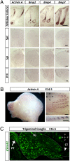
TGF-β ligand expression in the whisker pad and activation of TGF-β signaling in trigeminal sensory neurons. (A) In situ hybridization of Activin A, Bmp2, Bmp4, and Bmp7 in the whisker pad and in SpC, SpI, and PrV brainstem nuclei at E16.5. Dashed lines delineate the PrV nucleus. All transcripts are exclusively expressed in the whisker follicles. (B) Whole-mount in situ hybridization of Activin A at E14.5 reveals its expression in the whisker pad. Note the gradient established by the differential expression among whisker follicles along the A-P axis. (Lower Right) Magnified view of whisker row D. (C) pSmad2 immunolabeling of TG at E16.5 shows robust signal of pSmad2 in the cell bodies and axons of the PT (arrow) but not in the axons of the CT (arrowhead). CT, central tract. (Scale bars: A, 200 μm; B, 100 μm.)
Fig. 3.
Fig. 3.
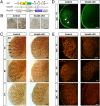
Smad4-cKO sensory neurons form defective barrelettes. (A) Schematic of the alleles used to generate [Advillincre/+; Smad4flox/flox] mice: AdvillinCre, Smad4flox, and ROSA26Sortm1(CAG-ALPP)Fawa (RosaPLAP). (B) In situ hybridization of Smad4 in Smad4-cKO mice at E15.5 confirms the deletion of Smad4 in trigeminal sensory neurons. Magnified view (Right) of the regions highlighted (Left). (C) CO staining of control and Smad4-cKO SpI nucleus at P1, P5, and P7. Note that at P1, barrelettes are distinguishable in control SpI (arrows) but imperceptible in Smad4-cKO SpI. At later stages, barrelette-like structures emerge in Smad4-cKO SpI, but defined barrelettes are never observed. (D) TenascinC immunostaining (green) of a P7 SpC nucleus. In control mice, barrelette boundaries are revealed by TenascinC (arrowheads), whereas in Smad4-cKO mice, this pattern is lost. (E) vGlut1 immunolabeling (red) of PrV, SpI, and SpC of P7 control and Smad4-cKO mice. vGlut1 staining reveals the emergence of barrelette-like structures in SpI and PrV nuclei and no barrelettes in the SpC nucleus of Smad4-cKO mice. (Scale bars: 100 μm; B, enlarged views, 25 μm.)
Fig. 4.
Fig. 4.
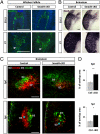
Smad4-cKO sensory neurons have normal peripheral innervation and central axon outgrowth but defective segregation of central projections. (A) PGP9.5 immunostaining of whisker follicles at E16.5 and P7 of control and Smad4-cKO mice. Sections were counterstained with DAPI. No difference in whisker sensory innervation is detected between control and Smad4-cKO mice at both time points. Arrowheads indicate longitudinal lanceolate endings at P7. (B) PLAP staining of sensory central projections to the SpI at E16.5 and P7 in control and Smad4-cKO mice. No major changes are detected. (C) Maximum projection images of confocal stacks depicting dextran-labeled central projections of control and Smad4-cKO sensory neurons innervating D2 and D3 whiskers in SpC (Upper) and SpI (Lower) nuclei at P3. Asterisks indicate centroids of D2 (blue) and D3 (yellow) corresponding barrelettes. Note that the relative orientation of the two centroids is affected by Smad4 ablation. (D) Average ± SEM of the percentage of overlap between labeled barrelettes in SpC and SpI (Ctrl: n = 3 mice, cKO: n = 6 mice). *P < 0.01; **P < 0.001. cKO, Smad4-cKO; Ctrl, control. (Scale bar: 100 μm.)
Fig. 5.
Fig. 5.
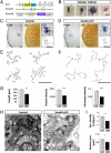
Atrophied terminal arborization and reduced number of synapses in Smad4-cKO sensory neurons. (A) Schematic of the alleles used here. CreERt2 was inserted into the Advillin locus to generate AdvillinCreERt2. The AdvillinCreERt2 line then was crossed with Smad4flox and the reporter line RosaPLAP. (B) PLAP staining of a single trigeminal mechanosensory neuron innervating one whisker follicle at P1. PLAP and CO staining of consecutive sections of control (C) and Smad4-cKO (D) SpI nuclei at P1. Magnified view (Upper Right) of the central terminal projection highlighted (Left). 2D projection (Lower Right) of the complete 3D Neurolucida reconstruction of the same terminal arbor. Representative 2D projections of terminal arbors in SpI nucleus of control (E) and Smad4-cKO (F) mice are shown. The dots (•) represent varicosities, defined as an enlargement of the axon. Note the decreased complexity and fewer varicosities present in Smad4-cKO neurons. (G) Quantitative analysis of total length and branch and varicosity densities of Ctrl and cKO terminal arbors (Ctrl: n = 47, four mice; cKO: n = 20, four mice). (H) EM images of control and Smad4-cKO terminal boutons of PrV nuclei at P7. White arrowheads indicate asymmetrical synaptic contacts, and red arrows indicate symmetrical contacts. Note the presence of fewer synapses in Smad4-cKO boutons. (I) Average ± SEM synaptic density (number of synapses/10 μm2) and bouton area (μm2) of cKO (n = 180 boutons) and Ctrl (n = 200 boutons) mice. *P < 0.05; **P < 0.01; ***P < 0.001 by the Student's t test. cKO, Smad4-cKO; Ctrl, control. (Scale bars: AD, 100 μm; CF, 50 μm in enlarged views; H, 500 nm.)

References

    1. Woolsey TA, Van der Loos H. The structural organization of layer IV in the somatosensory region (SI) of mouse cerebral cortex. The description of a cortical field composed of discrete cytoarchitectonic units. Brain Res. 1970;17:205–242. - PubMed
    1. Killackey HP, Rhoades RW, Bennett-Clarke CA. The formation of a cortical somatotopic map. Trends Neurosci. 1995;18:402–407. - PubMed
    1. Petersen CC. The functional organization of the barrel cortex. Neuron. 2007;56:339–355. - PubMed
    1. Erzurumlu RS, Murakami Y, Rijli FM. Mapping the face in the somatosensory brainstem. Nat Rev Neurosci. 2010;11:252–263. - PMC - PubMed
    1. Killackey HP, Fleming K. The role of the principal sensory nucleus in central trigeminal pattern formation. Brain Res. 1985;354:141–145. - PubMed

Publication types