. 2011 Feb 22;108(8):3246-51.
doi: 10.1073/pnas.1013225108. Epub 2011 Feb 7.

Low-resolution structure of a vesicle disrupting α-synuclein oligomer that accumulates during fibrillation

Affiliations

Low-resolution structure of a vesicle disrupting α-synuclein oligomer that accumulates during fibrillation

Lise Giehm et al. Proc Natl Acad Sci U S A. .

Abstract

One of the major hallmarks of Parkinson disease is aggregation of the protein α-synuclein (αSN). Aggregate cytotoxicity has been linked to an oligomeric species formed at early stages in the aggregation process. Here we follow the fibrillation process of αSN in solution over time using small angle X-ray scattering and resolve four major coexisting species in the fibrillation process, namely monomer, dimer, fibril and an oligomer. By ab initio modeling to fit the data, we obtain a low-resolution structure of a symmetrical and slender αSN fibril in solution, consisting of a repeating unit with a maximal distance of 900 Å and a diameter of ∼180 Å. The same approach shows the oligomer to be shaped like a wreath, with a central channel and with dimensions corresponding to the width of the fibril. The structure, accumulation and decay of this oligomer is consistent with an on-pathway role for the oligomer in the fibrillation process. We propose an oligomer-driven αSN fibril formation mechanism, where the fibril is built from the oligomers. The wreath-shaped structure of the oligomer highlights its potential cytotoxicity by simple membrane permeabilization. This is confirmed by the ability of the purified oligomer to disrupt liposomes. Our results provide the first structural description in solution of a potentially cytotoxic oligomer, which accumulates during the fibrillation of αSN.


Conflict of interest statement

The authors declare no conflict of interest.

Figures

Fig. 1.
Fig. 1.
Fibrillation of αSN above SCC reveals a sigmoidal curve and β-sheet formation. (A) Double logarithmic plot of the time required to reach 50% completion of the fibrillation versus the protein concentration. Linear regression yields two linear segments and the intercept between the lines defines SCC. Error bars represent the standard deviation with N = 3. (B) ThT-emission as a function of time, where each data point represents ThT-emission of a given sample that was subsequently used for SAXS solution measurements. The fibrillation experiments run for a total of 22.5 hr. (C) Far-UV CD spectrum of freshly prepared αSN (gray spheres) and mature fibrils after 22 hr (black spheres). (D) ATR-FTIR spectrum of mature fibrils after 22 hr. Both techniques confirm the appearance of extensive β-sheets.
Fig. 2.
Fig. 2.
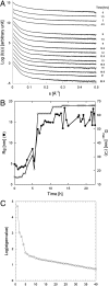
Analysis of SAXS data of the fibrillation process shows a time dependent increase in Rg and Dmax and SVD reveals four components. (A) Selected SAXS spectra showing the evolution of fibrillation from 0–22.5 hr in intervals of ∼1.5 hr. Curves are translated arbitrarily for viewing purposes. (B) Rg (black spheres) and Dmax (white spheres) plotted versus time. (C) Plot of the logarithm to the eigenvalues resulting from the singular value decomposition. The plot suggests 4 principal components.
Fig. 3.
Fig. 3.
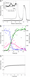
(A) Gel filtration chromatrogram of 12 mg/mL αSN at pH 7.4 (black spheres) and pH 10.5 (gray spheres). Insert shows zoom of oligomer peak at pH 7.4, which disappears at pH 10.5. (B) Development of the fibrillation process monitored by SAXS and ThT-emission. SAXS-derived volume fractions are plotted as a function of incubation time, illustrated by blue spheres (sum of monomer and dimer fractions), pink spheres (volume fraction of the fourth component), and green spheres (the volume fraction of fibril). The measured ThT-emission as a function of time is illustrated by black spheres. (C) The ability of purified αSN oligomers to permeabilise DOPG vesicles containing calcein. Black spheres illustrate the time profile of calcein release after addition of oligomers isolated from gel filtration experiments. The signal is normalized relative to signal from Triton-X induced complete rupture of the vesicles. As negative control we use monomer obtained by gel filtration (white spheres).
Fig. 4.
Fig. 4.
Low-resolution SAXS model of the oligomer, fibril, and an oligomer-driven fibril formation. (A) The P(r) of the early and late fibrils (large spheres, light and dark colors respectively), and the cross section of the early fibril (Insert, black spheres) compared with the P(r) of the oligomer [Insert, (white spheres)]. (B) SAXS-derived structure of the αSN oligomer. The average structure (mesh representation) and the filtered averaged structure (surface representation) are displayed and superimposed. The filtered structure has a volume corresponding to the average volume of individual models, and the difference between the average and filtered structure indicates the general level of differences between individual models. The model is showed in two orientations, rotated 90° around the longest axes. (C) The SAXS-derived model of the symmetric, early fibril that exists in solution in equilibrium with native species and a fourth component. A single repeating unit is shown in cyan, with the averaged model and the filtered averaged models superimposed in mesh-representation. The principle of the repeats building the mature fibrils is shown to the right, where three repeats of the filtrated model are shown. Repeats two and three have been translated 880 Å vertically with respect to the first repeat, and the model to the right is rotated 90° around a vertical axis with respect to the left model. (D) Model for the elongation of fibrils. In rose/purple colors, 26 oligomers constituting one repeating unit of the mature fibril (averaged and filtered model shown in cyan mesh) are displayed in surface representation. Below, the 26 oligomers are superimposed with the fibril repeating unit, whereas the two models are separated above. The lowest representation is rotated 90° around a horizontal axis with resepect to the top two models. Length scales are as in Fig. 4C.

Similar articles

Cited by

References

    1. Fink AL. The aggregation and fibrillation of alpha-synuclein. Acc Chem Res. 2006;39:628–634. - PubMed
    1. Volles MJ, Lansbury PT., Jr Vesicle permeabilization by protofibrillar alpha-synuclein is sensitive to Parkinson’s disease-linked mutations and occurs by a pore-like mechanism. Biochemistry. 2002;41:4595–4602. - PubMed
    1. Volles MJ, et al. Vesicle permeabilization by protofibrillar alpha-synuclein: implications for the pathogenesis and treatment of Parkinson’s disease. Biochemistry. 2001;40:7812–7819. - PubMed
    1. Conway KA, et al. Acceleration of oligomerization, not fibrillization, is a shared property of both alpha-synuclein mutations linked to early-onset Parkinson’s disease: implications for pathogenesis and therapy. Proc Natl Acad Sci USA. 2000;97:571–576. - PMC - PubMed
    1. Lashuel HA, et al. Alpha-synuclein, especially the Parkinson’s disease-associated mutants, forms pore-like annular and tubular protofibrils. J Mol Biol. 2002;322:1089–1102. - PubMed

Publication types

LinkOut - more resources