Ultra-high-field imaging distinguishes MS lesions from asymptomatic white matter lesions
- PMID: 21300968
- PMCID: PMC3053180
- DOI: 10.1212/WNL.0b013e31820b7630
Ultra-high-field imaging distinguishes MS lesions from asymptomatic white matter lesions
Abstract
Objectives: To investigate whether multiple sclerosis (MS) and non-MS white matter brain lesions can be distinguished by their appearance on 7 T T2*-weighted MRI.
Methods: This was an observational study of 28 patients with MS and 17 patients with cerebral white matter lesions who did not have MS. Subjects were imaged using 7 T T2*-weighted imaging. White matter lesions were identified and analyzed for volume, location, and perivenous appearance.
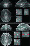
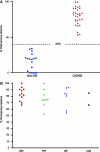
Results: Out of 901 lesions identified in patients with MS, 80% were perivenous. In comparison, 19% of 428 lesions identified in patients without MS had a perivenous appearance. Seven-Tesla T2*-weighted MRI reliably distinguished all patients with clinically definite MS (>40% lesions appeared perivenous) from those without clinical MS (<40% lesions appeared perivenous). Perivenous lesion appearance was more predictive of MS (odds ratio [OR] 14, p < 0.001) than subcortical or periventricular lesion location (OR 4.5, p < 0.001, and OR 2.4, p = 0.009). Perivenous lesion appearance was observed with a similar frequency in patients with clinically isolated syndrome of demyelination and in early (gadolinium-enhancing) MS lesions.
Conclusion: Perivenous lesion location on 7 T T2*-weighted imaging is predictive of the presence of demyelination. Optimization of this imaging technique at lower magnetic resonance field strengths would offer benefit for the diagnosis of MS.
Figures


Comment in
-
Multiple sclerosis: sun exposure and vitamin D, ultra-high-field imaging, and B cell clones in the meninges.J Neurol. 2011 Apr;258(4):708-10. doi: 10.1007/s00415-011-6000-6. J Neurol. 2011. PMID: 21431894 No abstract available.
References
-
- Polman CH, Reingold SC, Edan G, et al. Diagnostic criteria for multiple sclerosis: 2005 revisions to the “McDonald Criteria”. Ann Neurol 2005;58:840–846 - PubMed
-
- Newcombe J, Hawkins CP, Henderson CL, et al. Histopathology of multiple sclerosis lesions detected by magnetic resonance imaging in unfixed postmortem central nervous system tissue. Brain 1991;114:1013–1023 - PubMed
-
- Ormerod IE, Miller DH, McDonald WI, et al. The role of NMR imaging in the assessment of multiple sclerosis and isolated neurological lesions: a quantitative study. Brain 1987;110:1579–1616 - PubMed
-
- Liao D, Cooper L, Cai J, et al. The prevalence and severity of white matter lesions, their relationship with age, ethnicity, gender, and cardiovascular disease risk factors: the ARIC Study. Neuroepidemiology 1997;16:149–162 - PubMed
-
- Shintani S, Shiigai T, Arinami T. Silent lacunar infarction on magnetic resonance imaging (MRI): risk factors. J Neurol Sci 1998;160:82–86 - PubMed
Publication types
MeSH terms
Grants and funding
LinkOut - more resources
Full Text Sources
Other Literature Sources
Medical
