. 2011 Feb;8(1):015003.
doi: 10.1088/1478-3975/8/1/015003. Epub 2011 Feb 7.

Micromechanics of human mitotic chromosomes

Affiliations

Micromechanics of human mitotic chromosomes

Mingxuan Sun et al. Phys Biol. 2011 Feb.

Abstract

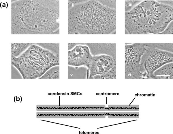
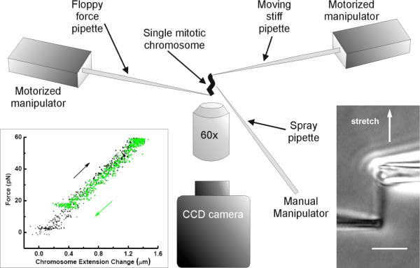
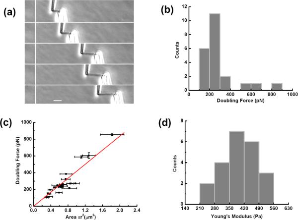
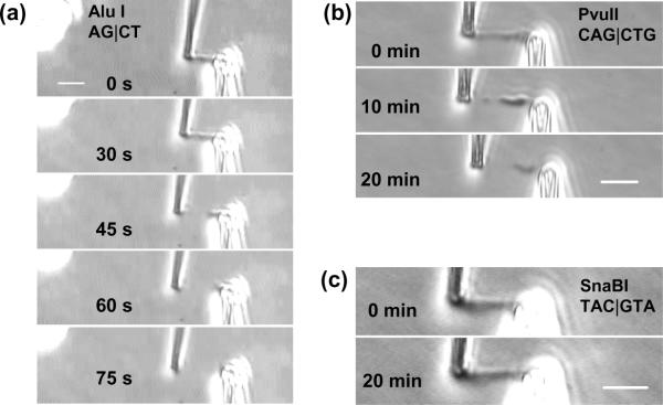
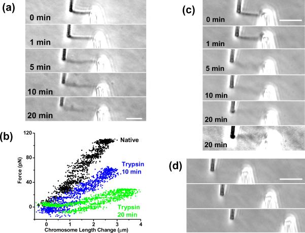
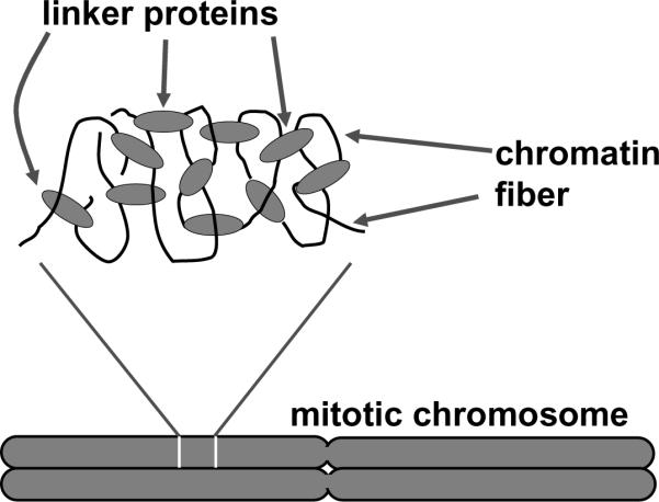
Eukaryote cells dramatically reorganize their long chromosomal DNAs to facilitate their physical segregation during mitosis. The internal organization of folded mitotic chromosomes remains a basic mystery of cell biology; its understanding would likely shed light on how chromosomes are separated from one another as well as into chromosome structure between cell divisions. We report biophysical experiments on single mitotic chromosomes from human cells, where we combine micromanipulation, nano-Newton-scale force measurement and biochemical treatments to study chromosome connectivity and topology. Results are in accord with previous experiments on amphibian chromosomes and support the 'chromatin network' model of mitotic chromosome structure. Prospects for studies of chromosome-organizing proteins using siRNA expression knockdowns, as well as for differential studies of chromosomes with and without mutations associated with genetic diseases, are also discussed.


Figures

Figure 1
Figure 1
Mitotic chromosome organization. (a) Cell cycle (newt Notophthalmus viridescens lung epithelial cell) showing interphase (i); prophase (ii); metaphase (iii); anaphase (iv); telophase (v); and interphase following division of daughter cells (vi) (pictures courtesy M. Poirier). Our chromosome-stretching experiments are carried out at “prometaphase”, just before capture of the mitotic chromosomes by the mitotic “spindle” apparatus (iii). Bar is 20 μm. (b) Sketch of metaphase chromosome showing side-by-side chromatids and centromere; chromatid-axial condensin distibution inferred from experiments [4, 7] are indicated by black dots.
Figure 2
Figure 2
Chromosome stretching experiment. A single mitotic chromosome following its removal from a dividing cell is suspended between two pipettes on motorized manipulators in the aqueous tissue culture medium in which the cells are grown. Inset at lower right shows a phase-contrast micrograph of chromosome and pipettes. Bar is 5 μm. Inset at lower left shows force-extension data for a human chromosome (black: extension, gray: retraction) showing its linear, reversible elasticity.
Figure 3
Figure 3
Human chromosomes display linear elasticity over a wide range that can be described by a Young's modulus of ≈ 420 Pa. (a) Images of a human chromosome during a stretching experiment. Bar is 5 μm. (b) Distribution of force constants from a series of 22 human chromosome-stretching experiments. (c) Plot of force constant vs cross-sectional area for the measurements of (b); the data fit well to a straight line with slope 420 ± 20 Pa. (d) Distribution of stretching (Young) modulus measurements from the measurements of (b); note the tighter distribution compared to the force constant distribution of (b). Mean Young modulus is 400 ± 20 Pa.
Figure 4
Figure 4
Restriction enzyme digestion of a human mitotic chromosome. Bars are all 5 μm (a) Photos top to bottom show cleavage and dissolution of a chromosome between the pipettes over the first 75 sec of spraying a chromosome with the blunt-cutter AluI (AG|CT). Cumulative enzyme spray time is indicated in each image. (b) Photos top to bottom show partial digestion of a chromosome by the 6-base cutter PvuII (CAG|CTG) after 20 min spraying. (c) Photos show no visible effect of the 6-base cutter SnaBI (TAC|GTA) on a human chromosome after 20 min spraying.
Figure 5
Figure 5
Effect of protein digestion on a human mitotic chromosome. Bars are all 5 μm. (a) Series of photos during digestion by trypsin showing gradual loss of phase contrast, lengthening, and thickening; cumulative enzyme spray time is indicated in each image. (b) Force-extension curves for the experiment of (a) showing that the chromosome remains elastic during the digestion. (c) Series of photos of mitotic chromosome during a digestion by proteinase K. The last two images show the same digestion time point (20 min); final image has higher contrast. Bar is 5 μm. (d) Series of photos showing the digested chromosome of (c) subjected to a stretching experiment indicating that it still supports stress.
Figure 6
Figure 6
“Chromatin network” model of mitotic chromosome folding. Chromatin segments (black curves) are connected together by protein-chromatin linkers (gray ovals). Cutting DNA (chromatin) disconnects the network, while cutting protein only lengthens and softens the network.
Figure 7
Figure 7
“X-shaped” human chromosome attached to pipettes. (a) Relaxed chromosome; the two-chromatid structure of the chromosome is evident in the image. Bar is 5 μm (b) Stretched chromosome; the left pipette is attached to the upper chromatid and is extending it more than the lower chromatid.

Similar articles

Cited by

References

    1. Hirano T. Biochemical and genetic dissection of mitotic chromosome condensation. Trends Biol. Sci. 1995;20:357–61. - PubMed
    1. Marko JF, Siggia ED. Polymer models of meiotic and mitotic chromosomes. Mol. Biol. Cell. 1997;8:2217–31. - PMC - PubMed
    1. Shintomi K, Hirano T. Sister chromatid resolution: a cohesin releasing network and beyond. Chromosoma. 2010;119:459–67. - PubMed
    1. Maeshima K, Laemmli UK. A two-step scaffolding model for mitotic chromosome assembly. Dev. Cell. 2003;4:467–80. - PubMed
    1. Kireeva N, Lakonishok M, Kireev I, Hirano T, Belmont AS. Visualization of early chromosome condensation: a hierarchical folding, axial glue model of chromosome structure. J. Cell Biol. 2004;166:775–85. - PMC - PubMed

Publication types

Substances

LinkOut - more resources