

Knock-down of amphiregulin inhibits cellular invasion in inflammatory breast cancer
- PMID: 21302279
- PMCID: PMC3865809
- DOI: 10.1002/jcp.22620
Knock-down of amphiregulin inhibits cellular invasion in inflammatory breast cancer
Abstract
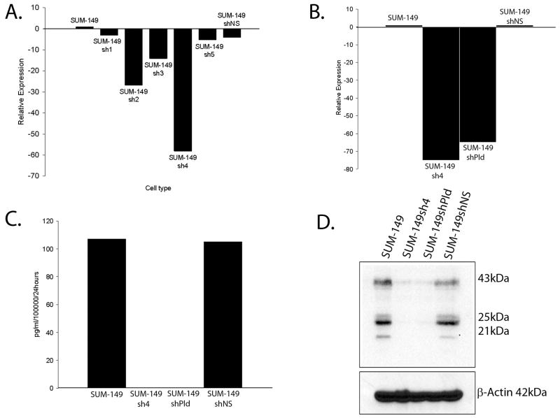
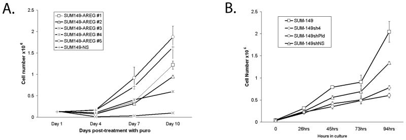
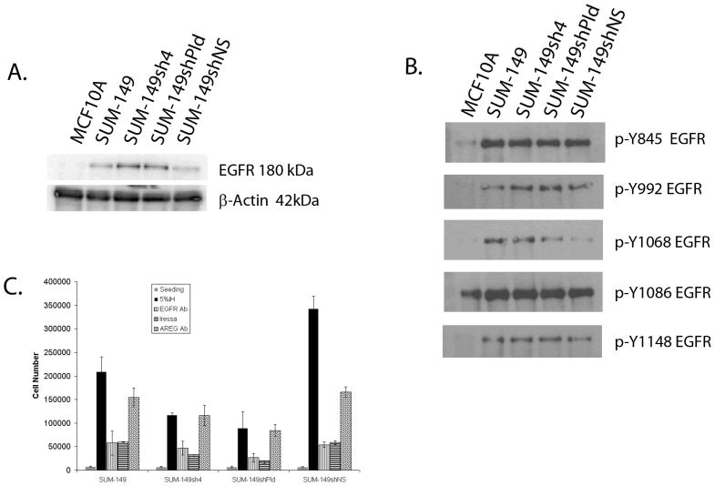
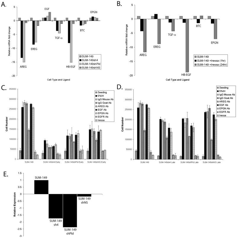
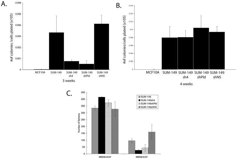
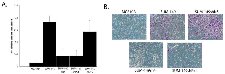
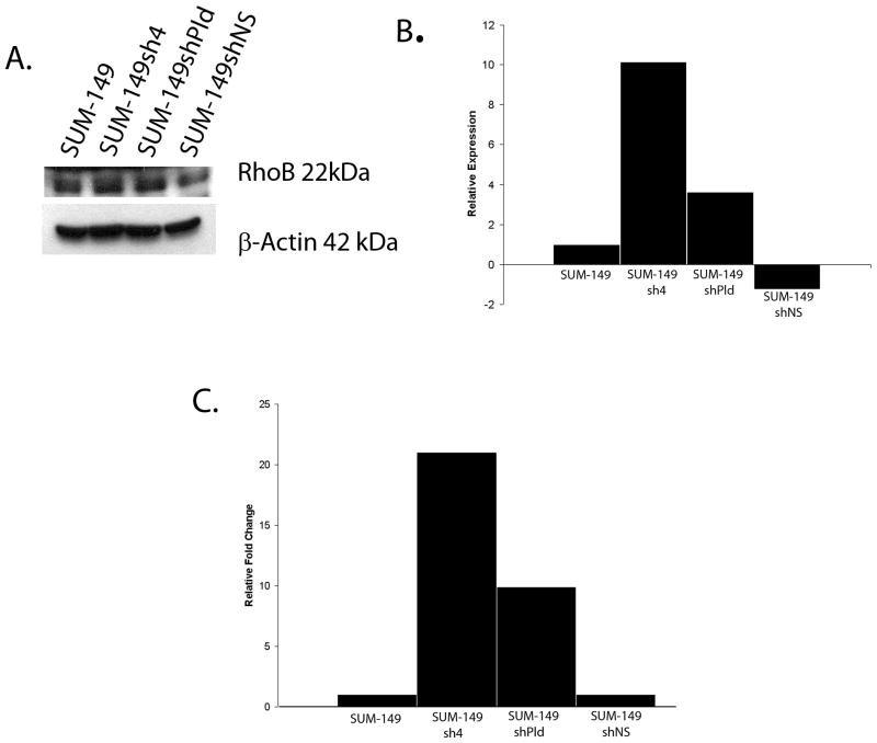
We have previously shown that SUM-149 human breast cancer cells require an amphiregulin (AREG) autocrine loop for cell proliferation. We also demonstrated that AREG can increase epidermal growth factor receptor (EGFR) stability and promote EGFR localization to the plasma membrane. In the present studies we successfully knocked-down AREG expression in SUM-149 cells by lentiviral infection of AREG shRNA. In the absence of AREG expression, SUM-149 cell growth was slowed, but not completely inhibited. Furthermore, cells infected with AREG shRNA constructs showed an increase in EGFR protein expression by Western blot. Immunofluorescence and confocal microscopy showed that following AREG knock-down, EGFR continued to localize to the cell surface. Soft agar assays demonstrated that AREG knock-down cells retain anchorage-independent growth capacity. Additionally mammosphere forming assays and Adefluor staining analysis showed that knock-down of AREG expression did not affect the expression of stem cell phenotypes. However, following AREG knock-down, SUM-149 cells demonstrated a dramatic decrease in their ability to invade a Matrigel matrix. Consistent with this observation, microarray analysis comparing cells infected with a non-silencing vector to the AREG knock-down cells, identified genes associated with the invasive phenotype such as RHOB and DKK1, and networks associated with cell motility such as integrin-linked kinase signaling, and focal adhesion kinase signaling. AREG was also found to modulate WNT and Notch signaling in these cells. Thus, AREG functions in regulating the invasive phenotype, and we propose that this regulation may be through altered signaling that occurs when AREG activates plasma membrane localized EGFR.
Copyright © 2011 Wiley-Liss, Inc.
Figures
Similar articles
-
Reconstitution of amphiregulin-epidermal growth factor receptor signaling in lung squamous cell carcinomas activates PTHrP gene expression and contributes to cancer-mediated diseases of the bone.Mol Cancer Res. 2009 Oct;7(10):1714-28. doi: 10.1158/1541-7786.MCR-09-0131. Epub 2009 Oct 13. Mol Cancer Res. 2009. PMID: 19825997 Free PMC article.
-
The role of amphiregulin in exemestane-resistant breast cancer cells: evidence of an autocrine loop.Cancer Res. 2008 Apr 1;68(7):2259-65. doi: 10.1158/0008-5472.CAN-07-5544. Cancer Res. 2008. PMID: 18381432 Free PMC article.
-
Cross-suppression of EGFR ligands amphiregulin and epiregulin and de-repression of FGFR3 signalling contribute to cetuximab resistance in wild-type KRAS tumour cells.Br J Cancer. 2012 Apr 10;106(8):1406-14. doi: 10.1038/bjc.2012.103. Br J Cancer. 2012. PMID: 22491422 Free PMC article.
-
The multiple roles of amphiregulin in human cancer.Biochim Biophys Acta. 2011 Dec;1816(2):119-31. doi: 10.1016/j.bbcan.2011.05.003. Epub 2011 May 30. Biochim Biophys Acta. 2011. PMID: 21658434 Review.
-
Amphiregulin as a novel target for breast cancer therapy.J Mammary Gland Biol Neoplasia. 2008 Jun;13(2):171-9. doi: 10.1007/s10911-008-9081-9. Epub 2008 Apr 25. J Mammary Gland Biol Neoplasia. 2008. PMID: 18437539 Review.
Cited by
-
Molecular signature induced by RNASET2, a tumor antagonizing gene, in ovarian cancer cells.Oncotarget. 2011 Jun;2(6):477-84. doi: 10.18632/oncotarget.274. Oncotarget. 2011. PMID: 21646684 Free PMC article. Review.
-
The molecular consequences of androgen activity in the human breast.Cell Genom. 2023 Mar 8;3(3):100272. doi: 10.1016/j.xgen.2023.100272. eCollection 2023 Mar 8. Cell Genom. 2023. PMID: 36950379 Free PMC article.
-
Oncogenic signaling in amphiregulin and EGFR-expressing PTEN-null human breast cancer.Mol Oncol. 2015 Feb;9(2):527-43. doi: 10.1016/j.molonc.2014.10.006. Epub 2014 Oct 23. Mol Oncol. 2015. PMID: 25454348 Free PMC article.
-
CD133 Stimulates Cell Proliferation via the Upregulation of Amphiregulin in Melanoma.Cells. 2024 May 2;13(9):777. doi: 10.3390/cells13090777. Cells. 2024. PMID: 38727313 Free PMC article.
-
Reporters to mark and eliminate basal or luminal epithelial cells in culture and in vivo.PLoS Biol. 2018 Jun 20;16(6):e2004049. doi: 10.1371/journal.pbio.2004049. eCollection 2018 Jun. PLoS Biol. 2018. PMID: 29924804 Free PMC article.
References
-
- Adnane J, Muro-Cacho C, Mathews L, Sebti SM, Munoz-Antonia T. Suppression of rho B expression in invasive carcinoma from head and neck cancer patients. Clin Cancer Res. 2002;8(7):2225–2232. - PubMed
-
- Berquin IM, Pang B, Dziubinski ML, Scott LM, Chen YQ, Nolan GP, Ethier SP. Y-box-binding protein 1 confers EGF independence to human mammary epithelial cells. Oncogene. 2005;24(19):3177–3186. - PubMed
-
- Brown CL, Meise KS, Plowman GD, Coffey RJ, Dempsey PJ. Cell surface ectodomain cleavage of human amphiregulin precursor is sensitive to a metalloprotease inhibitor. Release of a predominant N-glycosylated 43-kDa soluble form. J Biol Chem. 1998;273(27):17258–17268. - PubMed
-
- Charafe-Jauffret E, Ginestier C, Iovino F, Tarpin C, Diebel M, Esterni B, Houvenaeghel G, Extra JM, Bertucci F, Jacquemier J, Xerri L, Dontu G, Stassi G, Xiao Y, Barsky SH, Birnbaum D, Viens P, Wicha MS. Aldehyde dehydrogenase 1-positive cancer stem cells mediate metastasis and poor clinical outcome in inflammatory breast cancer. Clin Cancer Res. 2010;16(1):45–55. - PMC - PubMed
Publication types
MeSH terms
Substances
Grants and funding
LinkOut - more resources
Full Text Sources
Other Literature Sources
Medical
Molecular Biology Databases
Research Materials
Miscellaneous
