. 2011 Nov;226(11):2965-78.
doi: 10.1002/jcp.22644.

Tiam1 is recruited to β1-integrin complexes by 14-3-3ζ where it mediates integrin-induced Rac1 activation and motility

Affiliations

Tiam1 is recruited to β1-integrin complexes by 14-3-3ζ where it mediates integrin-induced Rac1 activation and motility

Timothy E O'Toole et al. J Cell Physiol. 2011 Nov.

Abstract

14-3-3 is an adaptor protein that localizes to the leading edge of spreading cells, returning to the cytoplasm as spreading ceases. Previously, we showed that integrin-induced Rac1 activation and spreading were inhibited by sequestration of 14-3-3ζ and restored by its overexpression. Here, we determined whether 14-3-3 mediates integrin signaling by localizing a guanine nucleotide exchange factor (GEF) to Rac1-activating integrin complexes. We showed that GST-14-3-3ζ recruited the Rac1-GEF, Tiam1, from cell lysates through Tiam1 residues 1-182 (N(1-182) Tiam1). The physiological relevance of this interaction was examined in serum-starved Hela cells plated on fibronectin. Both Tiam1 and N(1-182) Tiam1 were recruited to 14-3-3-containing β1-integrin complexes, as shown by co-localization and co-immunoprecipitation. Integrin-induced Rac1 activation was inhibited when Tiam1 was depleted with siRNA or by overexpression of catalytically inactive N(1-182) Tiam1, which was incorporated into 14-3-3/β1-integrin complexes and inhibited spreading in a manner that was overcome by constitutively active Rac1. Integrin-induced Rac1 activation, spreading, and migration were also inhibited by overexpression of 14-3-3ζ S58D, which was unable to recruit Tiam1 from lysates, co-immunoprecipitate with Tiam1, or mediate its incorporation into β1-integrin complexes. Taken together, these findings suggest a previously unrecognized mechanism of integrin-induced Rac1 activation in which 14-3-3 dimers localize Tiam1 to integrin complexes, where it mediates integrin-dependent Rac1 activation, thus initiating motility-inducing pathways. Moreover, since Tiam1 is recruited to other sites of localized Rac1 activation through its PH-CC-EX domain, the present findings show that a mechanism involving its N-terminal 182 residues is utilized to recruit Tiam1 to motility-inducing integrin complexes.


Figures

Fig. 1.
Fig. 1.
Identification of Tiam1 as a GEF recruited by GST-14–3-3ζ. GST-14–3-3ζ variants were quantified by Coomassie-blue staining of SDS gels (A) and incubated with platelet (B) or Hela cell (C) lysates. Proteins pulled down by the GST-14–3-3ζ beads or GST-beads alone were analyzed by Western blotting using Vav1 orTiam1 (C-16) antibodies. D: The specificity of theTiam1 antibodies was verified on Western blots ofNIH3T3 and Hela cell lysates by probing Western blots with antibodies in the absence (left-hand part) or presence (right-hand part) of a 100-fold excess ofC-16 immunizing peptide.
Fig. 2.
Fig. 2.
Tiam1 interacts with 14–3-3ζ through its N-terminal region. A: Schematic representation of Tiam1 and fragments utilized in pull-down assays. PH denotes Pleckstrin homology domain; CC, coiled-coil region; EX, extended structure; PDZ, PSD-95/DlgA/ZO-1 domain; and Dbl, Dbl homologydomain.B:CHOcellsweretransientlytransfectedwiththeindicatedFlag-taggedTiam1fragments.Thetopblotshowsexpressionlevels of Flag-tagged fragments in comparable amounts of CHO cell lysates; the middle blot shows Flag-tagged fragments recruited by GST-14–3-3ζ beads; the bottom blot shows the absence of Flag-tagged fragments in material recruited by control GST-beads.
Fig. 3.
Fig. 3.
Adhesion-induced co-immunoprecipitation ofTiam1, 14–3-3, and integrin. A: Platelets were lysed in suspension or allowed to spread on fibrinogen for the indicated times prior to lysis. Lysates were immunoprecipitated with 14–3-3ζ antibodies or control IgG and immunoprecipitates analyzed on Western blots probed with antibodies against 14–3-3ζ, Tiam1, and β3-integrin. B: Hela cells were cotransfected with Flag-tagged N1–415 Tiam1 fragment and either HA-tagged 14–3-3ζ or non-Tiam1-recruiting 14–3-3ζ S58D. Lysates were immunoprecipitated with antibodies against Flagepitope or isotype-specific control IgG. Western blots of immunoprecipitates were probed with antibodies against Flag-epitope, pan-14–3-3, and β1-integrin.
Fig. 4.
Fig. 4.
14–3-3 and Tiam1 localize to regions of extending membrane in spreading Hela cells. A: Hela cells were serum-starved and plated on fibronectin (Fn) or poly-L-lysine (PLL). At intervals, cells were lysed and levels of active Rac1 determined. The fibronectin and poly-L-lysine samples were run on the same gel but irrelevant intermediate lanes were deleted. B: Cells that were spreading on fibronectin were fixed and stained for actin filaments and Tiam1. Tiam1 localized to regions of extending membrane, which were identified by the presence of submembranous actin filaments. C: Tiam1 co-localized with 14–3-3 at the leading edge of spreading cells. In (B,C), examples of extending membrane are indicated with arrows. D: Cells were fixed 2 h after they were plated on fibronectin. At this time, spreading had ceased and both 14–3-3 and Tiam1 were absent from the membrane. Examples of focal adhesions are indicated with arrow heads; neither protein localized to these sites of integrin-mediated adhesion. Bars, 10μm.
Fig. 5.
Fig. 5.
14–3-3 and Tiam1 translocate to the leading edge in migrating Hela cells. Confluent monolayers of Hela cells were wounded, allowed to migrate into the wound for 5 h, fixed, and stained for actin filaments, β1-integrin, and either 14–3-3ζ (A) or Tiam1 (B). Examples of leading edge regions are shown with squares and regions at which stress fibers terminate at focal adhesions with rectangles. Co-localization of 14–3-3ζ (A) or Tiam (B) with β1 integrin was assessed by quantification of Pearson’s coefficient (Manders et al., 1993). Values are shown in bar graphs as mean±SEM. An asterisk indicates a significant difference between Pearson’s coefficient at the leading edge and that at other membrane regions, P < 0.0001. Bars, 10μm.
Fig. 6.
Fig. 6.
Tiam1 generates active Rac1 during β1-integrin-mediated spreading. A: Western blot of a representative experiment in which non-transfected Hela cells (C) or cells transfected with Tiam1-specific (si) or non-specific siRNA (NS) were plated on fibronectin in the absence of serum for 15min. The top blot shows Tiam1 levels. The lower blots show levels of active and total Rac1 in lysates of the corresponding cells. B: Levels of active Rac1 were normalized for total Rac1 and those in transfected cells expressed as a percentage of those in non-transfected cells. Data from three separate experiments were pooled; values shown are mean±SEM. The asterisk indicates a significant difference between levels in transfected compared to non-transfected cells, P<0.05.
Fig. 7.
Fig. 7.
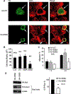
Inhibition of focal complex functions by a non-Tiam1-binding 14–3-3ζ variant. A: Hela cells were transiently transfected with HA-tagged wild-type (WT) 14–3-3ζ, Tiam1-binding S58A 14–3-3ζ, or non-Tiam1-binding S58D14–3-3ζ Cells were serum-starved, plated on fibronectin for2 h, fixed, and stained with HA antibodies and phalloidin. Arrows indicate cells expressing HA-14–3-3ζ proteins. Bar, 10μm. B: The mean area of non-expressing cells and cells expressing HA-14–3-3ζ proteins was quantified 60 min after plating on fibronectin; mean±SEM values for the indicated number of cells are shown. An asterisk indicates a significant difference between non-expressing cells (Con) and those expressing an HA-14–3-3ζ construct, P < 0.0003. Similar results were obtained in four different experiments for HA-WT and HA-S58D 14–3-3ζ and two different experiments for HA-S58A 14–3-3ζ C: Cells expressing comparable amounts of HA-wild-type, S58A, or S58D 14–3-3ζ were allowed to migrate across fibronectin-coated Transwell inserts for 1 and 4 h. Cells that migrated to the lower surface were quantified. For each bar, values shown represent the mean±SEM of six samples pooled from two different experiments. An asterisk indicates a significant difference between cells expressing wild-type 14–3-3ζ and a 14–3-3ζ variant, P < 0.02. D: Cells expressing comparable amounts of HA-S58A or HA-S58D 14–3-3ζ were plated on fibronectin for 10 min, lysed, and levels of total and active Rac1 determined. The left-hand part shows Western blots from a representative experiment. The top blot shows expression levels of Tiam1-binding 14–3-3ζ S58A and non-Tiam1-binding 14–3-3ζ S58D. The lower blots show levels of active and total Rac1 in the corresponding lysates. In the right-hand part, active Rac1 was normalized for total Rac1 and the level in 14–3-3ζ S58D-transfected cells expressed as a percentage of that in cells transfected with the Tiam-binding 14–3-3ζ S58A control. Mean±SEM values from three different experiments are shown. An asterisk indicates a significant difference between cells expressing non-Tiam1-binding 14–3-3ζ S58D and Tiam1-binding S58A control, P < 0.004.
Fig. 8.
Fig. 8.
Inhibition of focal complex functions by a 14–3-3-binding fragment of Tiam1.A: Hela cells that had been transiently transfected with Flag-tagged N1–182Tiam1 fragment were plated on fibronectin. At intervals, cells were fixed and actin filaments stained with phalloidin, Tiam1 fragment with Flag antibodies, and endogenous 14–3-3 with pan 14–3-3 antibodies. The upper part shows cells fixed 1 h after plating. In contrast to the N1–182 Tiam1-expressing cell indicated with an arrow, non-transfected cells were well spread. In the lower part, a representative transfected cell, fixed at 2 h, is shown at higher magnification. The N-terminal Tiam1 fragment and endogenous 14–3-3 continued to localize with β1-integrin at the periphery of the cell, which remained relatively unspread even 2 h after plating. Bars, 10μm. B: Cells were transiently transfected and plated on fibronectin for 1 h. In the left-hand graph, the mean area of 89 non-expressing and 77 Flag-taggedN1–182 Tiam1-expressing cells was quantified. In the right-hand graph, the mean area of 80 non-expressing and 20 Flag-tagged N182–415 Tiam1-expressing cells was quantified. Bar graphs show mean±SEM. An asterisk indicates a significant difference between expressing and non-expressing cells, P < 0.001. C: Cells that had been co-transfected with Flag-tagged N1–182 Tiam1 and HA-Q61LRac1 were serum-starved, plated on fibronectin for 1 h, fixed, and stained for actin filaments, Flag-tagged N1–182 Tiam1 (green), and HA-Q61LRac1 (cyan). The inhibitory effect of N1–182 Tiam1 on spreading was rescued by co-expression of Q61LRac1. Bar, 10μm.D: Hela cells that had been transiently transfected with vector or Flag-taggedTiam1 fragments were serum-starved, plated on fibronectin for 10min, lysed, and levels of total and active Rac1 determined. In the left hand part, Western blots from a representative experiment show expression of Tiam1 fragments, levels of active Rac1, and levels of total Rac1inthe corresponding samples. In the right-hand part, levels of active Rac1 were normalized and expressed as a percentage of that in cells transfected with vector alone. Values shown in the bar graph are the mean±SEM from four different experiments. An asterisk indicates a significant difference between cells transfected with a Tiam1 fragment and those transfected with vector, P < 0.05.

Similar articles

Cited by

References

    1. Aitken A 2006. 14–3-3 proteins: A historic overview. Semin Cancer Biol 16:162–172. - PubMed
    1. Aitken A, Baxter H, Dubois T, Clokie S, Mackie S, Mitchell K, Peden A, Zemlickova E. 2002. Specificity of14–3-3 isoform dimer interactions and phosphorylation. Biochem Soc Trans 30:351–360. - PubMed
    1. Albiges-Rizo C, Frachet P, Block MR. 1995. Down regulation of talin alters cell adhesion and the processing of the α5β1 integrin. J Cell Sci 108:3317–3329. - PubMed
    1. Angrand PO, Segura I, Volkel P, Ghidelli S, Terry R, Brajenovic M, Vintersten K, Klein R, Superti-Furga G, Drewes G, Kuster B, Bouwmeester T, Acker-Palmer A. 2006. Transgenic mouse proteomics identifies new 14–3-3-associated proteins involved in cytoskeletal rearrangements and cell signaling. Mol Cell Proteomics 5:2211–2227. - PubMed
    1. Arthur WT, Quilliam LA, Cooper JA. 2004. Rap1 promotes cell spreading by localizing Rac guanine nucleotide exchange factors. J Cell Biol 167:111–122. - PMC - PubMed

Publication types

MeSH terms

Substances