

Combining registration and active shape models for the automatic segmentation of the lymph node regions in head and neck CT images
- PMID: 21302791
- PMCID: PMC3000861
- DOI: 10.1118/1.3515459
Combining registration and active shape models for the automatic segmentation of the lymph node regions in head and neck CT images
Abstract
Purpose: Intensity-modulated radiation therapy (IMRT) is the state of the art technique for head and neck cancer treatment. It requires precise delineation of the target to be treated and structures to be spared, which is currently done manually. The process is a time-consuming task of which the delineation of lymph node regions is often the longest step. Atlas-based delineation has been proposed as an alternative, but, in the authors' experience, this approach is not accurate enough for routine clinical use. Here, the authors improve atlas-based segmentation results obtained for level II-IV lymph node regions using an active shape model (ASM) approach.
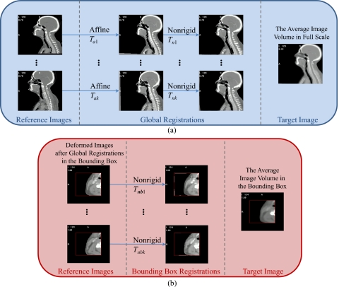
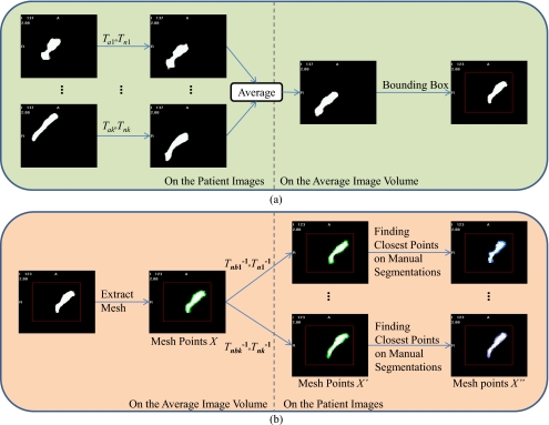
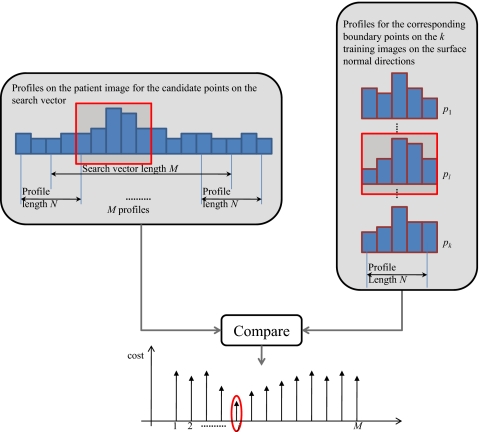
Methods: An average image volume was first created from a set of head and neck patient images with minimally enlarged nodes. The average image volume was then registered using affine, global, and local nonrigid transformations to the other volumes to establish a correspondence between surface points in the atlas and surface points in each of the other volumes. Once the correspondence was established, the ASMs were created for each node level. The models were then used to first constrain the results obtained with an atlas-based approach and then to iteratively refine the solution.
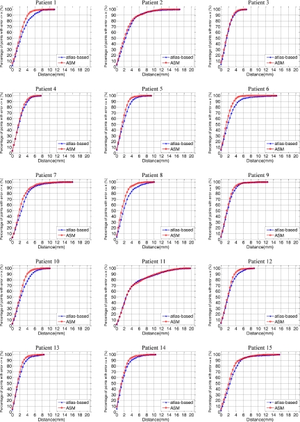
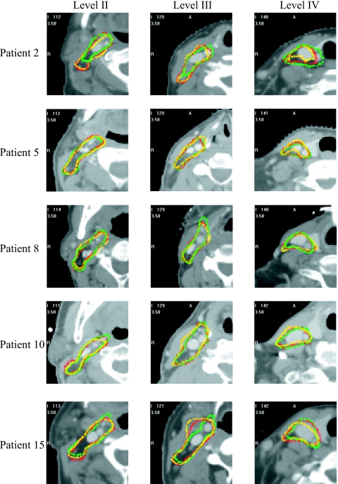
Results: The method was evaluated through a leave-one-out experiment. The ASM- and atlas-based segmentations were compared to manual delineations via the Dice similarity coefficient (DSC) for volume overlap and the Euclidean distance between manual and automatic 3D surfaces. The mean DSC value obtained with the ASM-based approach is 10.7% higher than with the atlas-based approach; the mean and median surface errors were decreased by 13.6% and 12.0%, respectively.
Conclusions: The ASM approach is effective in reducing segmentation errors in areas of low CT contrast where purely atlas-based methods are challenged. Statistical analysis shows that the improvements brought by this approach are significant.
Figures
References
-
- Commowick O., Warfield S. K., and Malandain G., “Using Frankenstein’s creature paradigm to build a patient specific atlas,” Proceedings of the 12th International Conference on Medical Image Computing and Computer Assisted Intervention (MICCAI ‘09), Vol. 5762, Pt. II, pp. 993–1000, September 2009. (unpublished). - PMC - PubMed
-
- Gorthi S., Duay V., Houhou N., Cuadra M. B., Schick U., Becker M., Allal A. S., and Thiran J. P., “Segmentation of head and neck lymph node regions for radiotherapy planning using active contour-based atlas registration,” IEEE J. Sel. Top. Signal Process. IJSTGY 3, 135–147 (2009).10.1109/JSTSP.2008.2011104 - DOI
-
- Cootes T. F., Taylor C. J., Cooper C. H., and Graham J., “Active shape models-their training and application,” Comput. Vis. Image Underst. CVIUF4 61, 38–59 (1995).10.1006/cviu.1995.1004 - DOI
Publication types
MeSH terms
Grants and funding
LinkOut - more resources
Full Text Sources
Medical
