

A comparative analysis of OTF, NPS, and DQE in energy integrating and photon counting digital x-ray detectors
- PMID: 21302803
- PMCID: PMC3016706
- DOI: 10.1118/1.3505014
A comparative analysis of OTF, NPS, and DQE in energy integrating and photon counting digital x-ray detectors
Abstract
Purpose: One of the benefits of photon counting (PC) detectors over energy integrating (EI) detectors is the absence of many additive noise sources, such as electronic noise and secondary quantum noise. The purpose of this work is to demonstrate that thresholding voltage gains to detect individual x rays actually generates an unexpected source of white noise in photon counters.
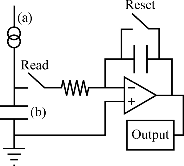
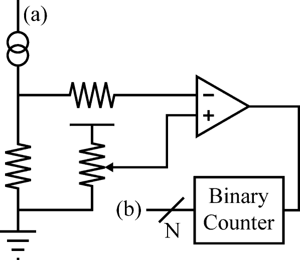
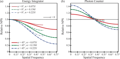
Methods: To distinguish the two detector types, their point spread function (PSF) is interpreted differently. The PSF of the energy integrating detector is treated as a weighting function for counting x rays, while the PSF of the photon counting detector is interpreted as a probability. Although this model ignores some subtleties of real imaging systems, such as scatter and the energy-dependent amplification of secondary quanta in indirect-converting detectors, it is useful for demonstrating fundamental differences between the two detector types. From first principles, the optical transfer function (OTF) is calculated as the continuous Fourier transform of the PSF, the noise power spectra (NPS) is determined by the discrete space Fourier transform (DSFT) of the autocovariance of signal intensity, and the detective quantum efficiency (DQE) is found from combined knowledge of the OTF and NPS. To illustrate the calculation of the transfer functions, the PSF is modeled as the convolution of a Gaussian with the product of rect functions. The Gaussian reflects the blurring of the x-ray converter, while the rect functions model the sampling of the detector.
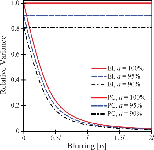
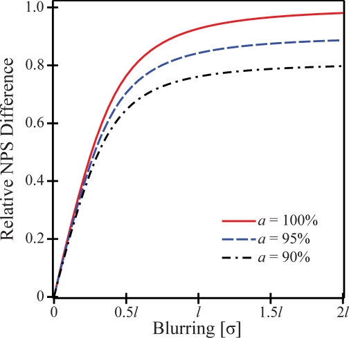
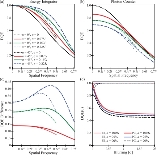
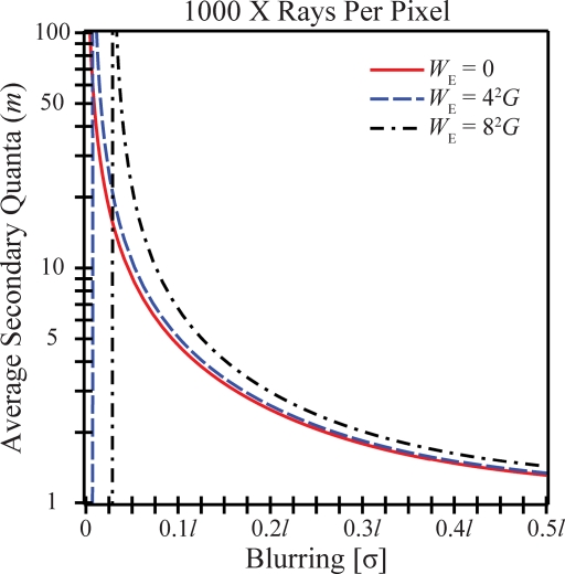
Results: The transfer functions are first calculated assuming outside noise sources such as electronic noise and secondary quantum noise are negligible. It is demonstrated that while OTF is the same for two detector types possessing an equivalent PSF, a frequency-independent (i.e., "white") difference in their NPS exists such that NPS(PC) > or = NPS(EI) and hence DQE(PC) < or = DQE(EI). The necessary and sufficient condition for equality is that the PSF is a binary function given as zero or unity everywhere. In analyzing the model detector with Gaussian blurring, the difference in NPS and DQE between the two detector types is found to increase with the blurring of the x-ray converter. Ultimately, the expression for the additive white noise of the photon counter is compared against the expression for electronic noise and secondary quantum noise in an energy integrator. Thus, a method is provided to determine the average secondary quanta that the energy integrator must produce for each x ray to have superior DQE to a photon counter with the same PSF.
Conclusions: This article develops analytical models of OTF, NPS, and DQE for energy integrating and photon counting digital x-ray detectors. While many subtleties of real imaging systems have not been modeled, this work is illustrative in demonstrating an additive source of white noise in photon counting detectors which has not yet been described in the literature. One benefit of this analysis is a framework for determining the average secondary quanta that an energy integrating detector must produce for each x ray to have superior DQE to competing photon counting technology.
Figures


Similar articles
-
A framework for performance characterization of energy-resolving photon-counting detectors.Med Phys. 2018 Nov;45(11):4897-4915. doi: 10.1002/mp.13172. Epub 2018 Oct 12. Med Phys. 2018. PMID: 30191571 Free PMC article.
-
An Analytical Model of NPS and DQE Comparing Photon Counting and Energy Integrating Detectors.Proc SPIE Int Soc Opt Eng. 2010 Feb;7622:76220I. doi: 10.1117/12.845310. Epub 2010 Mar 22. Proc SPIE Int Soc Opt Eng. 2010. PMID: 39086656 Free PMC article.
-
Spatial frequency-dependent pulse-height spectrum and method for analyzing detector DQE(f) from ensembles of single X-ray images.Med Phys. 2022 Jan;49(1):107-128. doi: 10.1002/mp.15344. Epub 2021 Nov 29. Med Phys. 2022. PMID: 34779519
-
Tutorial on X-ray photon counting detector characterization.J Xray Sci Technol. 2018;26(1):1-28. doi: 10.3233/XST-16210. J Xray Sci Technol. 2018. PMID: 29154310 Free PMC article. Review.
-
Photon-counting CT systems: A technical review of current clinical possibilities.Diagn Interv Imaging. 2025 Feb;106(2):53-59. doi: 10.1016/j.diii.2024.09.002. Epub 2024 Sep 20. Diagn Interv Imaging. 2025. PMID: 39304365 Review.
Cited by
-
Nonstationary model of oblique x-ray incidence in amorphous selenium detectors: II. Transfer functions.Med Phys. 2019 Feb;46(2):505-516. doi: 10.1002/mp.13312. Epub 2019 Jan 11. Med Phys. 2019. PMID: 30488455 Free PMC article.
-
A framework for performance characterization of energy-resolving photon-counting detectors.Med Phys. 2018 Nov;45(11):4897-4915. doi: 10.1002/mp.13172. Epub 2018 Oct 12. Med Phys. 2018. PMID: 30191571 Free PMC article.
-
Investigating the Potential for Super-Resolution in Digital Breast Tomosynthesis.Proc SPIE Int Soc Opt Eng. 2011 Feb;7961:79615K. doi: 10.1117/12.878845. Epub 2011 Mar 16. Proc SPIE Int Soc Opt Eng. 2011. PMID: 38846530 Free PMC article.
-
Detective quantum efficiency of photon-counting CdTe and Si detectors for computed tomography: a simulation study.J Med Imaging (Bellingham). 2020 Jul;7(4):043501. doi: 10.1117/1.JMI.7.4.043501. Epub 2020 Jul 17. J Med Imaging (Bellingham). 2020. PMID: 32715022 Free PMC article.
-
Optimization of phosphor-based detector design for oblique x-ray incidence in digital breast tomosynthesis.Med Phys. 2011 Nov;38(11):6188. doi: 10.1118/1.3639999. Med Phys. 2011. PMID: 22047384 Free PMC article.
References
-
- Rowlands J. A. and Yorkston J., “Flat panel detectors for digital radiography,” in Handbook of Medical Imaging Volume 1 Physics and Psychophysics, edited by Beutel J., Kundel H. L., and Van Metter R. L. (SPIE, Bellingham, 2000), Chap. 4, pp. 223–328.10.1117/3.832716.ch4 - DOI
-
- Jing T. et al., “Amorphous silicon pixel layers with cesium iodide converters for medical radiography,” IEEE Trans. Nucl. Sci. IETNAE 41(4), 903–909 (1994).10.1109/23.322829 - DOI
-
- Nagarkar V. V., Gupta T. K., Miller S. R., Klugerman Y., Squillante M. R., and Entine G., “Structured CsI(Tl) scintillators for x-ray imaging applications,” IEEE Trans. Nucl. Sci. IETNAE 45(3), 492–496 (1998).10.1109/23.682433 - DOI
Publication types
MeSH terms
Grants and funding
LinkOut - more resources
Full Text Sources
Other Literature Sources
