Comparative Study
. 2011 Jan;129(1):114-21.
doi: 10.1121/1.3523339.

Comparison between maximum radial expansion of ultrasound contrast agents and experimental postexcitation signal results

Affiliations
Comparative Study

Comparison between maximum radial expansion of ultrasound contrast agents and experimental postexcitation signal results

Daniel A King et al. J Acoust Soc Am. 2011 Jan.

Abstract

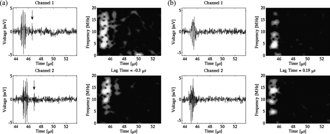
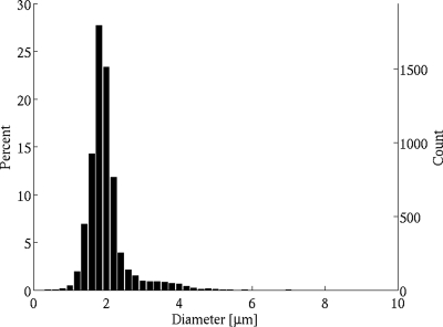
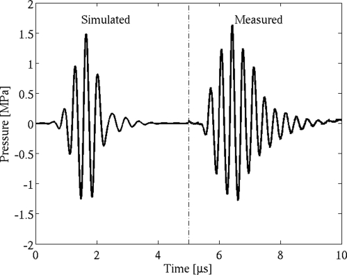
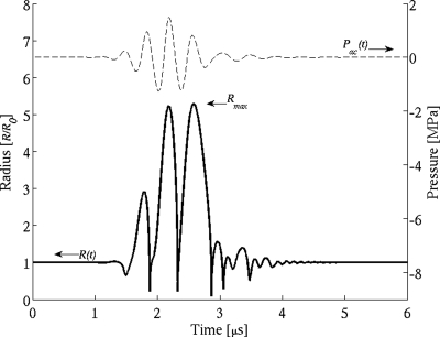
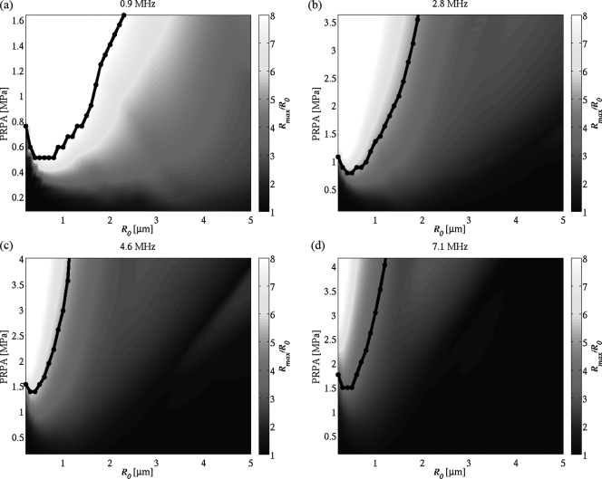
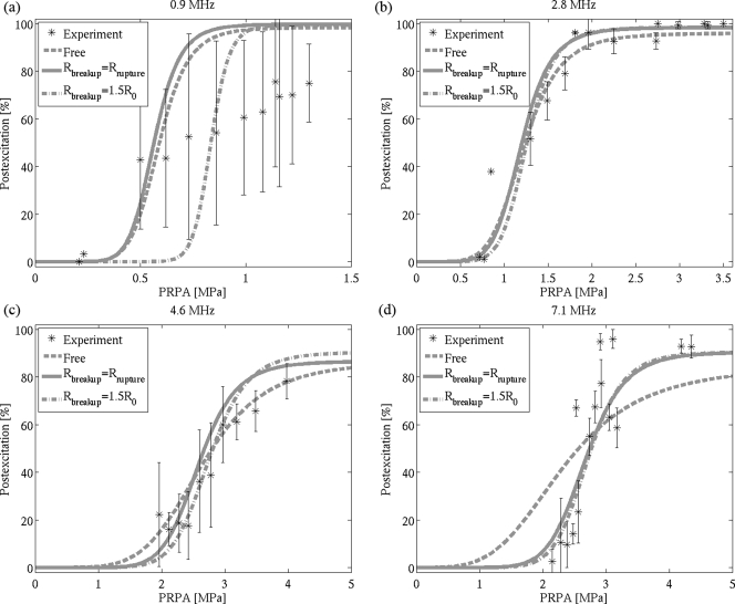
Experimental postexcitation signal data of collapsing Definity microbubbles are compared with the Marmottant theoretical model for large amplitude oscillations of ultrasound contrast agents (UCAs). After taking into account the insonifying pulse characteristics and size distribution of the population of UCAs, a good comparison between simulated results and previously measured experimental data is obtained by determining a threshold maximum radial expansion (Rmax) to indicate the onset of postexcitation. This threshold Rmax is found to range from 3.4 to 8.0 times the initial bubble radius, R0, depending on insonification frequency. These values are well above the typical free bubble inertial cavitation threshold commonly chosen at 2R0. The close agreement between the experiment and models suggests that lipid-shelled UCAs behave as unshelled bubbles during most of a large amplitude cavitation cycle, as proposed in the Marmottant equation.


Figures

Figure 1
Figure 1
(Color online) Examples of voltage–time signals (left columns) and their corresponding frequency–time images (right columns) from a Definity microbubble at 4.6 MHz and PRPA 4.47 MPa. (a) Single bubble with postexcitation (marked with an arrow) in both receive channels. (b) Single bubble with no postexcitation, only the principal response, in both receive channels.
Figure 2
Figure 2
Measured size distribution of approximately 6500 Definity microbubbles.
Figure 3
Figure 3
Simulated and measured three cycle, 2.8 MHz pulses (PRPA = 1.26 MPa).
Figure 4
Figure 4
Radial expansion of 1 μm radius Definity UCA for a 1.26 MPa PRPA pulse at 2.8 MHz.
Figure 5
Figure 5
(Color online) Maximum radial expansion Rmax of Definity bubbles calculated using the Marmottant equation with Rbreakup = Rrupture. The black lines indicate the threshold Rmax above which postexcitation is assumed to occur for optimal fit to the experimental results. (a) 0.9 MHz, threshold at 8.0R0; (b) 2.8 MHz, 5.0R0; (c) 4.6 MHz, 5.2R0; and (d) 7.1 MHz, 3.4R0.
Figure 6
Figure 6
Simulated percentage postexcitation curves for Definity using a free bubble model and the Marmottant model at (a) 0.9 MHz, (b) 2.8 MHz, (c) 4.6 MHz, and (d) 7.1 MHz. Experimental data points (mean ± standard deviation) are represented as asterisks (*).

References

    1. de Jong N., Emmer M., van Wamel A., and Versluis M., “Ultrasonic characterization of ultrasound contrast agents,” Med. Biol. Eng. Comput. 47, 861–873 (2009). 10.1007/s11517-009-0497-1 - DOI - PMC - PubMed
    1. Goldberg B. B., Raichlen J. S., and Forsberg F., Ultrasound Contrast Agents: Basic Principles and Clinical Applications, 2nd ed. (Martin Dunitz, London, 2001), pp. 1–440.
    1. Birnbaum Y., Luo H., Nagai T., Fishbein M. C., Peterson T. M., Li S., Kricsfeld D., Porter T. R., and Siegel R. J., “Noninvasive in vivo clot dissolution without a thrombolytic drug: Recanalization of thrombosed iliofemoral arteries by transcutaneous ultrasound combined with intravenous infusion of microbubbles,” Circulation 97, 130–134 (1998). - PubMed
    1. Datta S., Coussios C. C., Ammi A. Y., Mast T. D., de Courten-Myers G. M., and Holland C. K., “Ultrasound enhanced thrombolysis using definity as a cavitation nucleation agent,” Ultrasound Med. Biol. 34, 1421–1433 (2008). 10.1016/j.ultrasmedbio.2008.01.016 - DOI - PMC - PubMed
    1. McDannold N., Vykhodtseva N., and Hynynen K., “Targeted disruption of the blood–brain barrier with focused ultrasound: Association with cavitation activity,” Phys. Med. Biol. 51, 793–807 (2006). 10.1088/0031-9155/51/4/003 - DOI - PubMed

Publication types