. 2011 Apr;17(4):495-504.
doi: 10.1089/ten.tec.2010.0368. Epub 2011 Feb 8.

Noninvasive multimodal evaluation of bioengineered cartilage constructs combining time-resolved fluorescence and ultrasound imaging

Affiliations

Noninvasive multimodal evaluation of bioengineered cartilage constructs combining time-resolved fluorescence and ultrasound imaging

Brett Z Fite et al. Tissue Eng Part C Methods. 2011 Apr.

Abstract

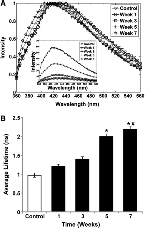
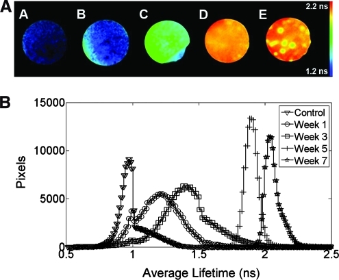
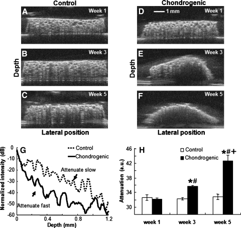
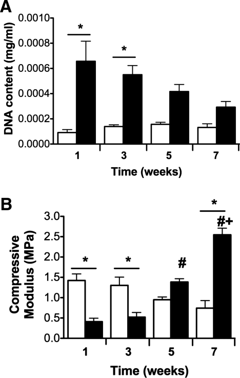
A multimodal diagnostic system that integrates time-resolved fluorescence spectroscopy, fluorescence lifetime imaging microscopy, and ultrasound backscatter microscopy is evaluated here as a potential tool for assessing changes in engineered tissue composition and microstructure nondestructively and noninvasively. The development of techniques capable of monitoring the quality of engineered tissue, determined by extracellular matrix (ECM) content, before implantation would alleviate the need for destructive assays over multiple time points and advance the widespread development and clinical application of engineered tissues. Using a prototype system combining time-resolved fluorescence spectroscopy, FLIM, and UBM, we measured changes in ECM content occurring during chondrogenic differentiation of equine adipose stem cells on 3D biodegradable matrices. The optical and ultrasound results were validated against those acquired via conventional techniques, including collagen II immunohistochemistry, picrosirius red staining, and measurement of construct stiffness. Current results confirm the ability of this multimodal approach to follow the progression of tissue maturation along the chondrogenic lineage by monitoring ECM production (namely, collagen type II) and by detecting resulting changes in mechanical properties of tissue constructs. Although this study was directed toward monitoring chondrogenic tissue maturation, these data demonstrate the feasibility of this approach for multiple applications toward engineering other tissues, including bone and vascular grafts.


Figures

FIG. 1.
FIG. 1.
(A) Normalized average fluorescence emission spectrum of chondrogenic samples retrieved for each time point (weeks 1, 3, 5, and 7) and control for comparison. Inset represents non-normalized emission spectra of the same samples. The spectra of all samples were averaged at each time point. (B) Average fluorescence lifetime averaged over all samples for each time point from TR-LIFS measurements (wavelength range 400–440 nm). The control value was obtained by averaging measurements of control samples for all time points. Data represent mean ± standard error of the mean (SEM) (n = 4, *p < 0.05 vs. control, #p < 0.05 vs. week 3).
FIG. 2.
FIG. 2.
(A) Representative average fluorescence lifetime images from fluorescence lifetime imaging microscopy measurements: (A) control sample and scaffolds cultured in chondrogenic conditions for (B) 1, (C) 3, (D) 5, and (E) 7 weeks. (B) Average fluorescence lifetime histogram of chondrogenic samples for each time point and control samples over 7 weeks (n = 4 for each week). Color images available online at www.liebertonline.com/tec.
FIG. 3.
FIG. 3.
Ultrasound backscatter microscopy images of (A–C) control scaffolds and (D–F) scaffolds cultured in chondrogenic conditions from week 1, 3, or 5 (top to bottom); (G) Changes in normalized intensity of the envelope of ultrasound signals as a function of sample depth; (H) changes in attenuation. Data are mean ± SEM from week 1 to 5 (*p < 0.05 vs. control sample at the same time point, #p < 0.05 vs. week 1, +p < 0.05 vs. weeks 1 and 3).
FIG. 4.
FIG. 4.
Biochemical staining for total collagen, collagen type II, and glycosaminoglycans. (A) Picrosirius red (PSR) staining of scaffolds cultured in chondrogenic conditions for 7 weeks and observed at 100 × magnification; scale bar represents 100 μm. Inset in upper right is PSR staining at 3 weeks in chondrogenic conditions, whereas inset in lower left is staining of construct in maintenance media after 7 weeks. (B) Quantification of collagen content as determined by PSR staining over 7 weeks; *p < 0.05 vs. control, #p < 0.05 vs. weeks 5 and 7, +p < 0.05 vs. all weeks). IHC for collagen type II after 3 (C) and 7 (D) weeks on construct in chondrogenic conditions; insets are constructs cultured in maintenance media for the same duration. Image shown at 10 × magnification; scale bar is 200 μm. Safranin-O stain of constructs in chondrogenic media after 3 (E) and 7 (F) weeks. Insets are constructs cultured in maintenance media for the same duration. Images shown at 40 × magnification; scale bar is 25 μm. Color images available online at www.liebertonline.com/tec.
FIG. 5.
FIG. 5.
(A) DNA content and (B) compressive moduli of cell-seeded constructs over time. Data are mean ± SEM (n = 4, *p < 0.05 vs. control sample at the same time point, #p < 0.05 vs. weeks 1 and 3, +p < 0.05 vs. weeks 1, 3, and 5). Control samples are open bars; chondrogenic samples are filled bars.
FIG. 6.
FIG. 6.
Linear regression of fluorescence intensity versus compressive modulus of chondrogenic samples (R2 = 0.998, p < 0.05) and versus total collagen content of chondrogenic samples (R2 = 0.981, p < 0.05). Square markers and dashed line correspond to collagen content (right) y-axis, whereas circular markers and solid line corresponds to compressive modulus (left) y-axis.

References

    1. Langer R. Vacanti J.P. Tissue engineering. Science. 1993;260:920. - PubMed
    1. Atala A. Engineering organs. Curr Opin Biotechnol. 2009;20:575. - PubMed
    1. Akhtar R. Schwarzer N. Sherratt M.J. Watson R.E. Graham H.K. Trafford A.W. Mummery P.M. Derby B. Nanoindentation of histological specimens: mapping the elastic properties of soft tissues. J Mater Res. 2009;24:638. - PMC - PubMed
    1. Ateshian G.A. Ricken T. Multigenerational interstitial growth of biological tissues. Biomech Model Mechanobiol. 2010;9:689. - PMC - PubMed
    1. Andersson-Engels S. Johansson J. Stenram U. Svanberg K. Svanberg S. Time-resolved laser-induced fluorescence spectroscopy for enhanced demarcation of human atherosclerotic plaques. J Photochem Photobiol B. 1990;4:363. - PubMed

Publication types

MeSH terms

LinkOut - more resources