

Expression and function of the autoimmune regulator (Aire) gene in non-thymic tissue
- PMID: 21303359
- PMCID: PMC3048612
- DOI: 10.1111/j.1365-2249.2010.04316.x
Expression and function of the autoimmune regulator (Aire) gene in non-thymic tissue
Abstract
Educational immune tolerance to self-antigens is induced primarily in the thymus where tissue-restricted antigens (TRAs) are presented to T lymphocytes by cells of the thymic stroma - a process known as central tolerance. The expression of these TRAs is controlled in part by a transcription factor encoded by the autoimmune regulatory (Aire) gene. Patients with a mutation of this gene develop a condition known as autoimmune-polyendocrinopathy-candidiasis-ectodermal-dystrophy (APECED), characterized by autoimmune destruction of endocrine organs, fungal infection and dental abnormalities. There is now evidence for TRA expression and for mechanisms of functional tolerance outside the thymus. This has led to a number of studies examining Aire expression and function at these extra-thymic sites. These investigations have been conducted across different animal models using different techniques and have often shown discrepant results. Here we review the studies of extra thymic Aire and discuss the evidence for its expression and function in both human and murine systems.
© 2011 The Authors. Clinical and Experimental Immunology © 2011 British Society for Immunology.
Figures
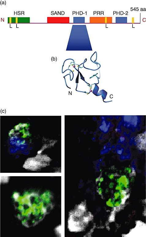
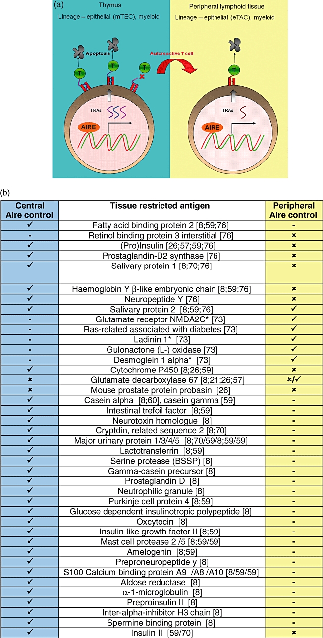
 : not done. (b) Comparison of thymic and peripheral Aire-restricted TRAs. *Human homologues of these mouse proteins have been described as autoantigens in human autoimmune diseases.
Similar articles
-
Normal thymic architecture and negative selection are associated with Aire expression, the gene defective in the autoimmune-polyendocrinopathy-candidiasis-ectodermal dystrophy (APECED).J Immunol. 2000 Aug 15;165(4):1976-83. doi: 10.4049/jimmunol.165.4.1976. J Immunol. 2000. PMID: 10925280
-
AIRE and immunological tolerance: insights from the study of autoimmune polyendocrinopathy candidiasis and ectodermal dystrophy.Curr Opin Allergy Clin Immunol. 2004 Dec;4(6):491-6. doi: 10.1097/00130832-200412000-00004. Curr Opin Allergy Clin Immunol. 2004. PMID: 15640689 Review.
-
APECED: A Paradigm of Complex Interactions between Genetic Background and Susceptibility Factors.Front Immunol. 2013 Oct 23;4:331. doi: 10.3389/fimmu.2013.00331. Front Immunol. 2013. PMID: 24167503 Free PMC article. Review.
-
Beyond APECED: An update on the role of the autoimmune regulator gene (AIRE) in physiology and disease.Autoimmun Rev. 2018 Apr;17(4):325-330. doi: 10.1016/j.autrev.2017.10.017. Epub 2018 Feb 8. Autoimmun Rev. 2018. PMID: 29427825 Review.
-
A defect of regulatory T cells in patients with autoimmune polyendocrinopathy-candidiasis-ectodermal dystrophy.J Immunol. 2007 Jan 15;178(2):1208-15. doi: 10.4049/jimmunol.178.2.1208. J Immunol. 2007. PMID: 17202386
Cited by
-
Neurotrophic keratitis in autoimmune polyglandular syndrome type 1: a case report.BMC Ophthalmol. 2021 Jan 7;21(1):17. doi: 10.1186/s12886-020-01770-w. BMC Ophthalmol. 2021. PMID: 33413189 Free PMC article.
-
Hematopoietic and stromal DMP1-Cre labeled cells form a unique niche in the bone marrow.Sci Rep. 2023 Dec 16;13(1):22403. doi: 10.1038/s41598-023-49713-x. Sci Rep. 2023. PMID: 38104230 Free PMC article.
-
Autoreactive T-Lymphocytes in Inflammatory Skin Diseases.Front Immunol. 2019 May 29;10:1198. doi: 10.3389/fimmu.2019.01198. eCollection 2019. Front Immunol. 2019. PMID: 31191553 Free PMC article. Review.
-
AIRE is induced in oral squamous cell carcinoma and promotes cancer gene expression.PLoS One. 2020 Feb 3;15(2):e0222689. doi: 10.1371/journal.pone.0222689. eCollection 2020. PLoS One. 2020. PMID: 32012175 Free PMC article.
-
A functional alternative splicing mutation in AIRE gene causes autoimmune polyendocrine syndrome type 1.PLoS One. 2013;8(1):e53981. doi: 10.1371/journal.pone.0053981. Epub 2013 Jan 8. PLoS One. 2013. PMID: 23342054 Free PMC article.
References
-
- Hiesche KD, Revesz L. Cellular repopulation in irradiated mouse thymus and bone marrow. Beitr Pathol. 1974;151:304–16. - PubMed
-
- Zlotoff DA, Schwarz BA, Bhandoola A. The long road to the thymus: the generation, mobilization, and circulation of T-cell progenitors in mouse and man. Semin Immunopathol. 2008;30:371–82. - PubMed
-
- Chien YH, Gascoigne NR, Kavaler J, Lee NE, Davis MM. Somatic recombination in a murine T-cell receptor gene. Nature. 1984;309:322–6. - PubMed
-
- Klein L, Hinterberger M, Wirnsberger G, Kyewski B. Antigen presentation in the thymus for positive selection and central tolerance induction. Nat Rev Immunol. 2009;9:833–44. - PubMed
Publication types
MeSH terms
Substances
LinkOut - more resources
Full Text Sources
