. 2011 Aug;51(8):1755-68.
doi: 10.1111/j.1537-2995.2010.03041.x. Epub 2011 Feb 8.

Fukuoka-1 strain of transmissible spongiform encephalopathy agent infects murine bone marrow-derived cells with features of mesenchymal stem cells

Affiliations

Fukuoka-1 strain of transmissible spongiform encephalopathy agent infects murine bone marrow-derived cells with features of mesenchymal stem cells

Larisa Cervenakova et al. Transfusion. 2011 Aug.

Abstract

Background: The possible risk of iatrogenic transmissible spongiform encephalopathies (TSEs, prion diseases) from transplantation of marrow-derived mesenchymal stem cells (MSCs) is uncertain. While most cell lines resist infection, a few propagate TSE agents.

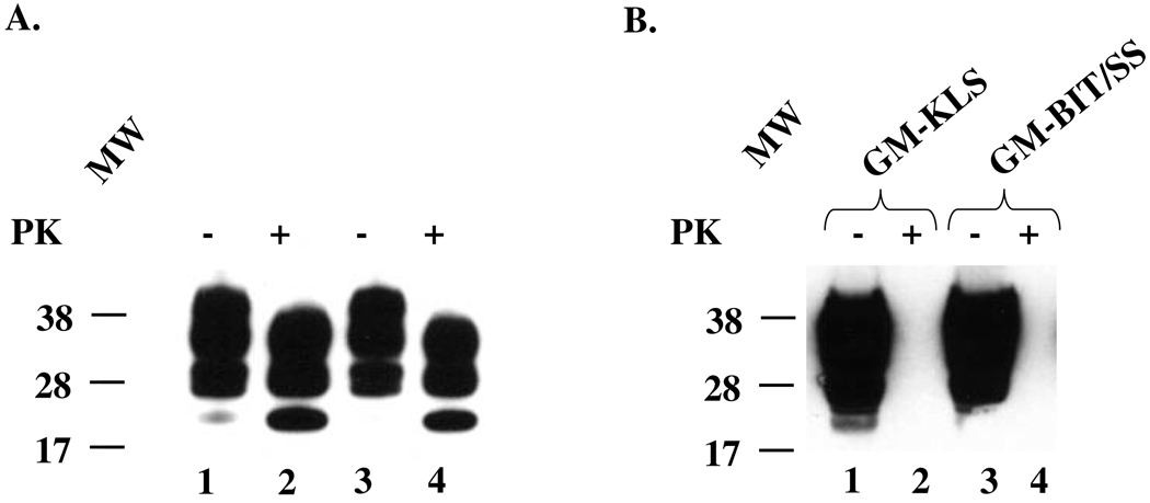
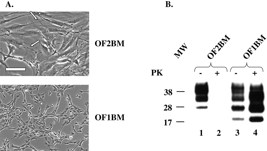
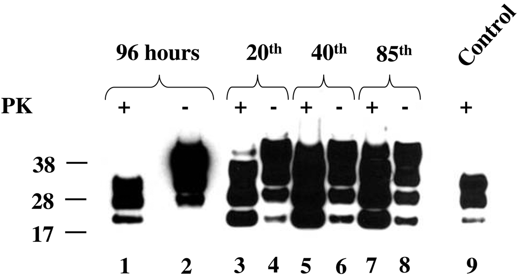
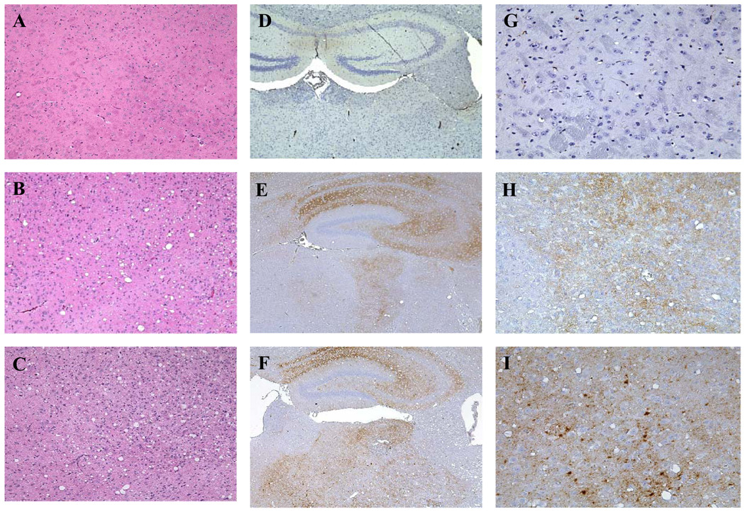
Study design and methods: We generated MSC-like (MSC-L) cell cultures from bone marrow (BM) of mice inoculated with the human-derived Fukuoka-1 (Fu) strain of TSE agent. Cultured cells were characterized for various markers and cellular prion protein (PrP(C) ) by fluorescence-activated cell sorting and for PrP(C) and its pathologic TSE-associated form (PrP(TSE) ) by Western blotting (WB). Cell cultures were tested for their susceptibility to infection with Fu in vitro. The infectivity of one Fu-infected cell culture was assayed in mice.

Results: BM cells from Fu-infected mice expressed neither PrP(C) nor PrP(TSE) after 3 days in culture as demonstrated by WB. Cells adherent to plastic and maintained under two different culture conditions became spontaneously immortalized and began to express PrP(C) at about the same time. One culture became transformed shortly after exposure to Fu in vitro and remained persistently infected, continuously generating PrP(TSE) through multiple passages; the infectivity of cultured cells was confirmed by intracerebral inoculation of lysates into mice. Both persistently TSE-infected and uninfected cells expressed a number of typical MSC markers.

Conclusion: BM-derived MSC-L cells of mice became persistently infected with the Fu agent under certain conditions in culture-conditions that differ substantially from those currently used to develop investigational human stem cell therapies.


Conflict of interest statement

Conflict of interest: The authors declare that they have no conflicts of interest relevant to the manuscript submitted to TRANSFUSION.

Figures

Fig. 1
Fig. 1. Immunoblots for PrP
(A). Total PrP (lanes 1 and 3) and PrPTSE (lanes 2 and 4) in samples of 0.1 % brain homogenates (10 µL loaded into the gel) of two SJL/Ola mice infected with Fukuoka-1 agent: lanes 1 and 3 and lanes 2 and 4 – samples untreated and treated with 100 µg/mL proteinase K (PK), respectively. The same two mice were used as sources of bone marrow (BM) described below. (B). BM-derived stromal cell cultures O1BM and O2BM (day 57 following isolation) maintained in two different growth media: GM-KLS or GM-BIT/SS media (see “Materials and Methods”). The presence of PrPC is evidenced by strong signals in PK-untreated samples (−) (lanes 1 and 3), and its absence in PK-treated samples (+) (lanes 2 and 4). The membranes were developed using the 6D11 antibody to PrP. A molecular mass standard in kilodaltons is shown on the left.
Fig. 2
Fig. 2. Characterization of BM stromal cell cultures
(A). Morphology. Untransformed OF2BM cells with fibroblast-like polygonal shapes, some visible stress fibers, and many well-developed filopodia (arrows). Transformed OF1BM cells showed morphological changes including a marked decrease in cell spreading, disappearance of stress fibers, and a smaller number of filopodia. Scale bar, 200 mm. (B). Immunoblot for PrP. PrPC (lane 1) but not PrPTSE (lane 2) is present in OF2BM cell lysate but both forms of PrP are present in OF1BM cell lysate (lane 3 and 4, respectively). Proteinase K (PK)-untreated samples (−) (lanes 1 and 3) and samples treated with 10 µg/mL PK (+) (lanes 2 and 4) Three glycoforms of PrP are displayed as bands corresponding to di-, mono-, and non-glycosylated isoforms. Note that the PK-treated samples of cell lysates were five-fold more concentrated than PK-untreated samples. The membrane was developed using 6D11 antibody. Molecular mass standard in kilodaltons is shown on the left.
Fig. 3
Fig. 3. Immunoblots for PrP from OF1BM cell cultures infected with Fukuoka-1 (Fu) agent
Cell lysates were prepared from cells collected at 96 hr (lanes 1 and 2) after in vitro exposure to Fu agent and from several subsequent serial passages (lanes 3 – 8). Presence of total PrP and PrPTSE is evidenced in samples untreated (−) or treated (+) with 10 µg/mL Proteinase K (PK), respectively. Note that the PK-treated samples (+) were five-fold more concentrated than samples not treated with PK (−). Control (lane 9) – sample of 0.1 % brain homogenate (10 µL loaded into the gel) treated with 100 µg/mL of PK. Three glycoforms of PrP are displayed as bands corresponding to di-, mono-, and non-glycosylated isoforms. The membranes were developed using 6D11 antibody. A molecular mass standard in kilodaltons is shown on the left.
Fig. 4
Fig. 4. Differentiation of OF1BM and OF2BM cells in cultures
OF1BM and OF2BM cell cultures frozen at passages 5 were recovered for the differentiation study. Controls represent cells maintained in medium without differentiation factors (Panels A, E, C, G). Adipogenic and osteogenic cultures were maintained in media with appropriate differentiation factors for 21 days (see “Materials and Methods”) (Panels B and F and D and H, respectively). Panels A, B, E, F – cells stained with 0.5% Oil Red O; panels C, D, G, H – cells stained with Alizarin Red S. Cells were fixed with either 10% formalin (panels A, B, I,J) or 70% ethanol (panels C, D, K, L) before staining. Original magnification, ×10.
Fig. 5
Fig. 5. Immunoblot comparing PrPTSE extracted from brain tissue of mice and from OF1BM cell lysate
Lane 1 – extract from the pooled brains of Swiss-Webster mice inoculated with Fukuoka-1 (Fu) agent; lane 2 – extract from lysate of an OF1BM cell culture persistently infected with Fu agent; lane 3 – extract from the brain of an FVB mouse inoculated with OF1BM cell lysate. All samples were treated with 100 µg/mL Proteinase K (PK). Three glycoforms of PrP are displayed as bands corresponding to di-, mono-, and non-glycosylated isoforms. The membranes were developed using 6D11 antibody. Molecular mass standard in kilodaltons is on the left.
Fig. 6
Fig. 6. Comparative histological features in brain tissues of FVB mice inoculated with OF1BM cell lysate or with brain homogenate infected with Fu agent
Panels A, D, G — representative sections of the cerebrum of a healthy mouse inoculated 570 days earlier with cells not exposed to Fukuoka-1 (Fu) agent: note absence of spongiform degeneration (A) and lack of PrP accumulation by immunostaining (D, G). Panels B, E, H — representative sections of the cerebrum of a sick mouse inoculated 114 days earlier with 4 × 10⁸ Fu-infected OF1BM cells, showing severe spongiform degeneration (B) and fine-punctate PrP-positive deposits (E, H). Panels C, F, I — representative sections of the cerebrum of a sick mouse 162 days after inoculation with a mouse brain homogenate (10−6 dilution) containing Fu agent, showing severe spongiform degeneration (C) and fine punctate and coarse PrP-positive deposits (F, I). Sections in panels A, B, C were stained with hematoxylin and eosin (original magnification, ×10). Sections in panels D, G, E, H, F, I were immunostained with anti-PrP antibody 6H4; D, E and F (original magnification, ×4). Sections in panels G, H, and I (original magnification, ×20).

Similar articles

Cited by

References

    1. Bruce ME, Will RG, Ironside JW, McConnell I, Drummond D, Suttie A, McCardle L, Chree A, Hope J, Birkett C, Cousens S, Fraser H, Bostock CJ. Transmissions to mice indicate that 'new variant' CJD is caused by the BSE agent. Nature. 1997;389:498–501. - PubMed
    1. Brown P, Brandel JP, Preece M, Sato M. Iatrogenic Creutzfeldt-Jakob disease: the waning of an era. Neurology. 2006;67:389–393. Epub (2006 Jul 19). Erratum in: Neurology 2006; 67:1528. Preese, Michael [corrected to Preece, Michael] - PubMed
    1. Hewitt PE, Llewelyn CA, Mackenzie J, Will RG. Creutzfeldt-Jakob disease and blood transfusion: results of the UK Transfusion Medicine Epidemiological Review study. Vox Sang. 2006;91:221–230. - PubMed
    1. Peden A, McCardle L, Head MW, Love S, Ward HJ, Cousens SN, Keeling DM, Millar CM, Hill FG, Ironside JW. Variant CJD infection in the spleen of a neurologically asymptomatic UK adult patient with haemophilia. Haemophilia. 2010;16:296–304. Epub 2010 Jan 12. - PubMed
    1. Dorsey K, Zou S, Schonberger KB, Sullivan M, Kessler D, Notari E, 4th, Fang CT, Dodd RY. Lack of evidence of transfusion transmission of Creutzfeldt-Jakob disease in a US surveillance study. Transfusion. 2009;49:977–984. Epub 2009 Jan 5. - PubMed

Publication types

MeSH terms