. 2011 Feb 8;2(1):2.
doi: 10.1186/2040-2392-2-2.

Neuropathologic features in the hippocampus and cerebellum of three older men with fragile X syndrome

Affiliations

Neuropathologic features in the hippocampus and cerebellum of three older men with fragile X syndrome

Claudia M Greco et al. Mol Autism. .

Abstract

Background: Fragile X syndrome (FXS) is the most common inherited form of intellectual disability, and is the most common single-gene disorder known to be associated with autism. Despite recent advances in functional neuroimaging and our understanding of the molecular pathogenesis, only limited neuropathologic information on FXS is available.

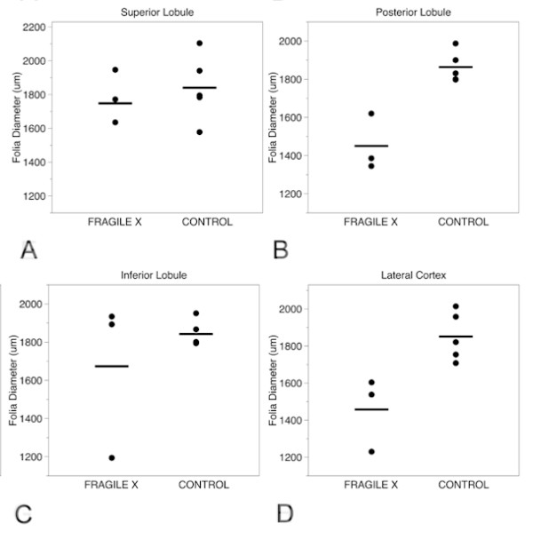
Methods: Neuropathologic examinations were performed on post-mortem brain tissue from three older men (aged 57, 64 and 78 years) who had received a clinical or genetic diagnosis of FXS. In each case, physical and cognitive features were typical of FXS, and one man was also diagnosed with autism. Guided by reports of clinical and neuroimaging abnormalities of the limbic system and cerebellum of individuals with FXS, the current analysis focused on neuropathologic features present in the hippocampus and the cerebellar vermis.

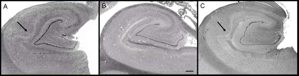
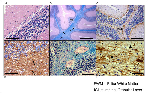
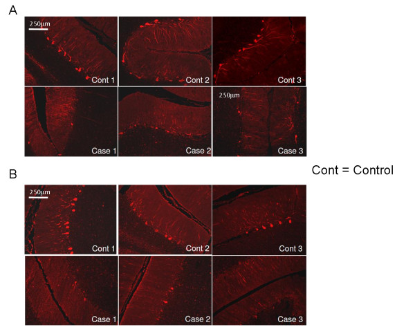
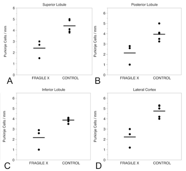
Results: Histologic and immunologic staining revealed abnormalities in both the hippocampus and cerebellar vermis. Focal thickening of hippocampal CA1 and irregularities in the appearance of the dentate gyrus were identified. All lobules of the cerebellar vermis and the lateral cortex of the posterior lobe of the cerebellum had decreased numbers of Purkinje cells, which were occasionally misplaced, and often lacked proper orientation. There were mild, albeit excessive, undulations of the internal granular cell layer, with patchy foliar white matter axonal and astrocytic abnormalities. Quantitative analysis documented panfoliar atrophy of both the anterior and posterior lobes of the vermis, with preferential atrophy of the posterior lobule (VI to VII) compared with age-matched normal controls.

Conclusions: Significant morphologic changes in the hippocampus and cerebellum in three adult men with FXS were identified. This pattern of pathologic features supports the idea that primary defects in neuronal migration, neurogenesis and aging may underlie the neuropathology reported in FXS.


Figures

Figure 1
Figure 1
Hippocampal formation. Images of the hippocampal formation at the level of the lateral geniculate body from (A) patient 1 and (C) patient 3 show abnormal expansion of CA1 by increased numbers of pyramidal neurons. These are compared with the more usual hippocampal microarchitecture that shows a thinner linear band of neurons in CA1, as seen in (B) a 62-year-old male control. Haematoxylin and eosin, original magnification ×10; scale bar = 1 mm. Arrow indicates bulge/expansion composed of increased numbers of pyramidal cells in (A) patient 1 and (C) patient 3.
Figure 2
Figure 2
Abnormalities in the cerebellum. Histologic abnormalities seen in FXS cerebella. (A) Decreased numbers of Purkinje cells (PCs) and clusters of PCs (arrows) in which cells are often misoriented. Haematoxylin and eosin, original magnification ×200; scale bar = 200 μm. (B) Patchy pallor of foliar white matter and increased variability in thickness of the internal granular cell layer (IGL). Arrows indicate foci of myelin pallor. Luxol fast blue-periodic-acid-Schiff (LFB-PAS), original magnification ×200; scale bar = 1 mm. (C) Diffuse axonal loss in cerebellar foliar white matter (FWM) (neurofilament immunohistochemistry, original magnification ×100; scale bar = 400 μm); (D) Activated astrocytes in foliar white matter, a finding not seen in deep cerebellar white matter. Arrows indicate abnormal astrocytes. (glial fibrillary acidic protein immunohistochemistry, original magnification ×200; scale bar = 200 μm). (E) Vascular hyalinosis within foliar white matter, as indicated by arrows. LFB-PAS, original magnification ×200; scale bar = 200 μm. (F) High magnification of foliar white matter axonal abnormalities that include axonal loss (arrowheads) and swollen axons, as indicated by arrows. Neurofilament immunohistochemistry, original magnification ×400; scale bar = 100 μm.
Figure 3
Figure 3
Calbindin immunofluorescent staining in the cerebellar cortex. Calbindin immunofluorescent staining of the cerebellar cortex indicates decreased numbers of PCs and diminished complexity of the PC dendritic arborization compared with age-matched controls. Similar changes were observed in all vermal lobules and in lateral posterior lobe cortex. (A) Vermal inferior lobule and (B) lateral posterior lobe cerebellar cortex. Original magnification ×10; scale bar = 250 μm.
Figure 4
Figure 4
Purkinje cell counts in the cerebellum. Purkinje cell (PC) counts in: (A) superior lobule; (B) posterior lobule; (C) inferior lobule; and (D) lateral cortex, in the three fragile X cases and age-matched controls.
Figure 5
Figure 5
Foliar diameter in the cerebellum. Foliar width measurements in: (A) superior lobule; (B) posterior lobule; (C) inferior lobule; and (D) lateral cortex, in the three fragile X cases and age-matched controls.

Similar articles

Cited by

References

    1. Belmonte MK, Bourgeron T. Fragile X syndrome and autism at the intersection of genetic and neural networks. Nat Neurosci. 2006;9:1221–1225. doi: 10.1038/nn1765. - DOI - PubMed
    1. Hagerman RJ, Rivera SM, Hagerman PJ. The fragile X family of disorders: A model for autism and targeted treatments. Curr Pediatr Rev. 2008;4:40–52. doi: 10.2174/157339608783565770. - DOI
    1. Kaufmann WE, Cortell R, Kau AS, Bukelis I, Tierney E, Gray RM, Cox C, Capone GT, Stanard P. Autism spectrum disorder in fragile X syndrome: communication, social interaction, and specific behaviors. Am J Med Genet. 2004;129A:225–234. doi: 10.1002/ajmg.a.30229. - DOI - PubMed
    1. Rogers SJ, Wehner EA, Hagerman RJ. The behavioral phenotype in fragile X: Symptoms of autism in very young children with fragile X syndrome, idiopathic autism, and other developmental disorders. J Dev Behav Pediatr. 2001;22:409–417. - PubMed
    1. Hatton DD, Sideris J, Skinner M, Mankowski J, Bailey DB Jr, Roberts JE, Mirrett P. Autistic behavior in children with fragile X syndrome: Prevalence, stability, and the impact of FMRP. Am J Med Genet A. 2006;140:1804–1813. - PubMed