

The functional neuroanatomy of dystonia
- PMID: 21303695
- PMCID: PMC3478782
- DOI: 10.1016/j.nbd.2011.01.026
The functional neuroanatomy of dystonia
Abstract
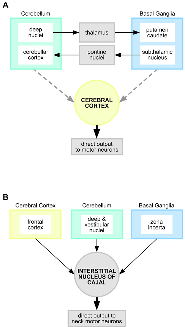
Dystonia is a neurological disorder characterized by involuntary twisting movements and postures. There are many different clinical manifestations, and many different causes. The neuroanatomical substrates for dystonia are only partly understood. Although the traditional view localizes dystonia to basal ganglia circuits, there is increasing recognition that this view is inadequate for accommodating a substantial portion of available clinical and experimental evidence. A model in which several brain regions play a role in a network better accommodates the evidence. This network model accommodates neuropathological and neuroimaging evidence that dystonia may be associated with abnormalities in multiple different brain regions. It also accommodates animal studies showing that dystonic movements arise with manipulations of different brain regions. It is consistent with neurophysiological evidence suggesting defects in neural inhibitory processes, sensorimotor integration, and maladaptive plasticity. Finally, it may explain neurosurgical experience showing that targeting the basal ganglia is effective only for certain subpopulations of dystonia. Most importantly, the network model provides many new and testable hypotheses with direct relevance for new treatment strategies that go beyond the basal ganglia. This article is part of a Special Issue entitled "Advances in dystonia".
Copyright © 2011 Elsevier Inc. All rights reserved.
Figures
References
-
- Alarcon F, et al. Focal limb dystonia in a patient with a cerebellar mass. Arch. Neurol. 2001;58:1125–1127. - PubMed
-
- Ali SO, et al. Alterations in CNS activity induced by botulinum toxin treatment in spasmodic dysphonia: an H215O PET study. J Speech Lang Hear Res. 2006;49:1127–1146. - PubMed
-
- Anheim M, et al. Ataxia with oculomotor apraxia type 2: clinical, biological and genotype/phenotype correlation study of a cohort of 90 patients. Brain. 2009;132:2688–2698. - PubMed
-
- Asanuma K, et al. Neuroimaging in human dystonia. J. Med. Investig. 2005a;52:272–279. - PubMed
Publication types
MeSH terms
Grants and funding
LinkOut - more resources
Full Text Sources
