. 2011 Apr;9(4):448-61.
doi: 10.1158/1541-7786.MCR-10-0471. Epub 2011 Feb 8.

Low-dose valproic acid enhances radiosensitivity of prostate cancer through acetylated p53-dependent modulation of mitochondrial membrane potential and apoptosis

Affiliations

Low-dose valproic acid enhances radiosensitivity of prostate cancer through acetylated p53-dependent modulation of mitochondrial membrane potential and apoptosis

Xufeng Chen et al. Mol Cancer Res. 2011 Apr.

Abstract

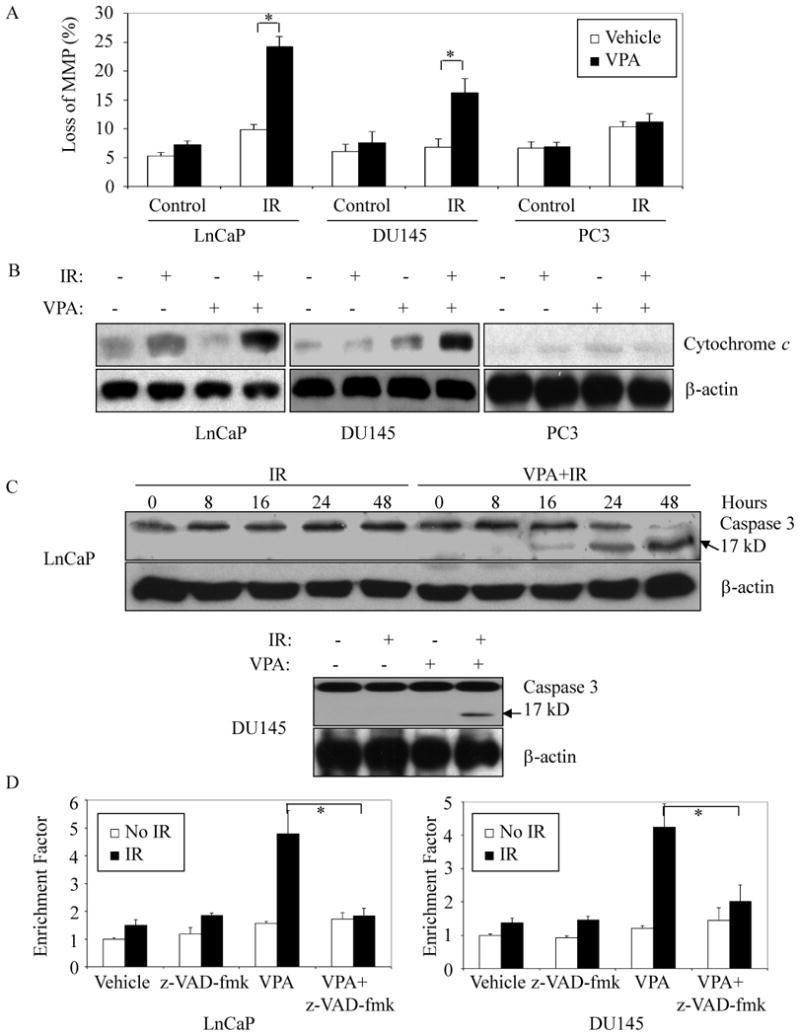
Histone deacetylase inhibitors (HDI) have shown promise as candidate radiosensitizers for many types of cancers, including prostate cancer. However, the mechanisms of action are not well understood. In this study, we show in prostate cancer cells that valproic acid (VPA) at low concentrations has minimal cytotoxic effects yet can significantly increase radiation-induced apoptosis. VPA seems to stabilize a specific acetyl modification (lysine 120) of the p53 tumor suppressor protein, resulting in an increase in its proapoptotic function at the mitochondrial membrane. These effects of VPA are independent of any action of the p53 protein as a transcription factor in the nucleus, since these effects were also observed in native and engineered prostate cancer cells containing mutant forms of p53 protein having no transcription factor activity. Transcription levels of p53-related or Bcl-2 family member proapoptotic proteins were not affected by VPA exposure. The results of this study suggest that, in addition to nuclear-based pathways previously reported, HDIs may also result in radiosensitization at lower concentrations via a specific p53 acetylation and its mitochondrial-based pathway(s).


Figures

Figure 1
Figure 1. VPA differentially induced radiosensitization and apoptosis in CaP cells
(A) Clonogenic survival assays of CaP cells pretreated with indicated doses of VPA. Log phase cells were trypsinized and plated as single cells. After 8 hours incubation to allow for cell attachment, cells were pretreated with different concentrations of VPA for 12 hours and then exposed to IR. Colony survival was determined 14-20 days later. (B) VPA increased apoptotic response to irradiation. Cells were first exposed to 50 μM VPA or left untreated for 12 hours before 10 Gy of IR. Cells were then collected 48 hours later, and apoptosis was evaluated by Annexin V-FITC apoptosis assay. Data represent the average of three to four experiments. Error bars indicate standard deviation. * indicates significant difference (ρ < 0.01).
Figure 2
Figure 2. Changes in mitochondrial membrane potential and caspase cascade activation
(A and B) VPA prior to irradiation increased radiation-induced mitochondrial membrane potential and cytochrome c release. Cells were pretreated with 50 μM VPA or left untreated for 12 hours followed by 10 Gy IR. Cells were then trypsinized and collected 24 hours later. Mitochondrial membrane potential was quantified by measurement of JC-1 fluorescence intensity (A); cytosolic protein was prepared as described in Materials and Methods, and analyzed by immunoblot to determine the release of cytochrome c from mitochondria. β-actin was included to confirm equivalent protein loadings (B). (C) VPA induced activation of caspase 3 in response to irradiation. Whole cell lysates were prepared 24 hours later, or at different time points as indicated after radiation. Immunoblots were performed to determine the activation of caspase-3. Cleaved caspase-3 presents product at 17kD. (D) Inhibition of VPA-effects on IR-induced apoptosis by the caspase inhibitor z-VAD-fmk. LnCaP or DU145 cells were pretreated with or without 50 μM VPA for 12 hours followed by 10 Gy IR. z-VAD-fmk (10 μM) was added to cells 2 hours before IR. Cells were maintained in VPA-containing medium for 48 hours. 1×10⁵ cells were then collected and lysed, and cell lysates were subjected to ELISA analysis as per manufacturer's instructions (Cell Death Detection ELISAPLUS). Error bars indicate one standard deviation from three to four individual experiments. * indicates significant difference (ρ < 0.01).
Figure 3
Figure 3. Mitochondrial accumulations of p53 and Bcl-2 family membrane proteins
(A) LnCaP cells were treated with IR (10 Gy) alone or with 50 μM VPA for 12 hours prior to IR. Mitochondrial fractions were prepared at indicated times and subjected to immunoblot using anti-p53 antibody (DO-1). Anti β-actin, or mt-HSP70 antibodies were included to confirm equivalent protein loading, respectively. (B) LnCaP, DU145 and PC-3 cells were pretreated with or without 50 μM VPA for 12 hours before 10 Gy of IR was delivered. Mitochondrial fractions (top) and whole cell lysates (bottom) were then prepared 24 hours later. p53, Bcl-2, Bcl-xL and Bax levels were analyzed by immunoblot. mt-Hsp70 was included to show equivalent protein loading. The purity of mitochondrial fractions was assessed by immunoblot with anti-cytochrome c, HSP70 and PCNA, a cytosolic/nuclear protein proliferating cell nuclear antigen (right panel). CF: cytosolic fraction; MF: mitochondrial fraction. The data are representative of three independent experiments.
Figure 4
Figure 4. Requirement of p53 in VPA-enhanced apoptotic response to IR
LnCaP cells were transiently transfected with p53 SiRNA oligos to reduce protein level of p53 (A), and pretreated with or without 50 μM VPA for 12 hours before IR (10 Gy). Cells were collected 48 hours after irradiation, and annexin V-FITC apoptosis assay was performed to analyze apoptosis (B). Mitochondrial and cytosolic fractions were prepared and analyzed by Immunoblot using anti-Bax, Bcl-xL and cytochrome c antibodies. β-actin and mt-HSP70 were included to confirm equivalent protein loading (C). Data represent the average of three experiments. Error bars indicate one standard deviation. * indicates significant difference (ρ < 0.01).
Figure 5
Figure 5. Engineering expression of wild-type p53 or mutant p53223Leu, but not mutant p53274Phe, restores the ability of VPA to enhance radiation-induced apoptotic response in p53 null PC-3 cells
PC-3 cells were stably transfected with wild-type p53, mutant p53223Leu, mutant p53274Phe or empty vector as control (A), and pretreated with or without 50 μM VPA for 12 hours before IR (10 Gy). For apoptosis analysis, cells were collected 48 hours later, and analyzed by annexin V-FITC apoptosis assay (B). Whole cell lysates or mitochondrial fraction were also prepared 24 hours after IR, and analyzed by immunoblot. β-actin and mt-HSP70 were included to confirm equivalent protein loading (C). Data represent the average of three experiments. Error bars indicate one standard deviation from three individual experiments. * and ** indicate significant differences (ρ < 0.01 or 0.05).
Figure 6
Figure 6. Loss of p53 acetylation at K120 reduced VPA-enhanced apoptosis response and radiosensitization
(A) VPA induced acetylation of mitochondrial p53 at K120 in response to irradiation. LnCaP cells were pretreated with 50 μM VPA for 12 hours and then irradiated (10Gy). The mitochondrial fraction was isolated 8 hours later, and subjected to immunoprecipitation with ac-K120-p53 antiserum. The resultant proteins were analyzed by immunoblot with p53 antibody (DO-1). Total p53 and mt-HSP70 were included to confirm equivalent protein loading; (B) Mutation at K120 of p53 (K120R) diminished VPA-enhanced loss of mitochondrial membrane potential (MMP) in response to irradiation in PC-3 cells engineered to express wild-type p53 or mutant p53223Leu. Stable transfectants of PC-3 cells with empty vector, wild-type p53, and mutant p53 (p53K120R, p53223Leu, p53274Phe, p53223Leu+K120R, and p53274Phe+K120R) were pretreated with VPA for 12 hours, and then irradiated (10Gy). Cells were collected by trypsinization 48 hours later, and stained with JC-1. Loss of MMP was analyzed by flow cytometry assay. Data represent the average of three experiments. Error bars indicate one SD from three individual experiments.* indicates significant difference (ρ < 0.01).
Figure 7
Figure 7. In Vitro cytochrome c release assay and acetylation assay
(A and B) Engineered PC-3 cells with empty vector, p53wt, p53K120R, p53223L or p53274P were treated with 50 μM VPA, irradiation or the combination as described above. Five hours later after the delivery of 10 Gy of IR, cytoplasmic extracts were prepared, and incubated with purified mitochondrial fraction from HCT116/p53−/− cells at 30°C for 40 min. The supernatant fraction and the pellets, after centrifugation at 12000 g for 10 min, were analyzed by immunoblots. (C) Purified p53 protein was incubated with Tip60, or BSA as control at 30°C for one hour, and the supernatant was directly analyzed for acetylation of K120 or p53, or incubated with purified mitochondria for cytochrome c release. (D) DU145 cells were engineered for wt-Tip60 expression, and pretreated with 50 μM VPA or 200 nM SAHA, followed by 10 Gy of irradiation. Cells were analyzed for apoptosis 48 hours later by determining the enrichment of mono- and oligonucleosomes in the cytoplasm using ELISA assay. Error bars indicate one SD from three individual experiments.
Figure 8
Figure 8. VPA induces radiosensitization in prostate cancer in vivo
Athymic nude mice bearing isogenic DU145 xenograft tumors were treated with VPA (300mg/kg × 6) administered intraperitoneally every 12 hours for 3 days and/or 10 Gy IR. In the combined treatment group, IR was delivered after the third injection of VPA. The growth curves represent the average value in each group of 5-8 mice. Error bars represent one standard error (S.E.).

References

    1. Howell SB. Resistance to apoptosis in prostate cancer cells. Mol Urol. 2000;4:225–229. discussion 231. - PubMed
    1. Zietman AL, DeSilvio ML, Slater JD, Rossi CJ, Jr, Miller DW, Adams JA, Shipley WU. Comparison of conventional-dose vs high-dose conformal radiation therapy in clinically localized adenocarcinoma of the prostate: a randomized controlled trial. Jama. 2005;294:1233–1239. - PubMed
    1. Pollack A, Zagars GK, Starkschall G, Antolak JA, Lee JJ, Huang E, von Eschenbach AC, Kuban DA, Rosen I. Prostate cancer radiation dose response: results of the M. D. Anderson phase III randomized trial. Int J Radiat Oncol Biol Phys. 2002;53:1097–1105. - PubMed
    1. Zelefsky MJ, Fuks Z, Hunt M, Lee HJ, Lombardi D, Ling CC, Reuter VE, Venkatraman ES, Leibel SA. High dose radiation delivered by intensity modulated conformal radiotherapy improves the outcome of localized prostate cancer. J Urol. 2001;166:876–881. - PubMed
    1. Camphausen K, Tofilon PJ. Inhibition of histone deacetylation: a strategy for tumor radiosensitization. J Clin Oncol. 2007;25:4051–4056. - PubMed

Publication types

MeSH terms