. 2011 Apr 8;286(14):12743-55.
doi: 10.1074/jbc.M110.199737. Epub 2011 Feb 8.

Potent and selective inhibitors of the inositol-requiring enzyme 1 endoribonuclease

Affiliations

Potent and selective inhibitors of the inositol-requiring enzyme 1 endoribonuclease

Kori Volkmann et al. J Biol Chem. .

Abstract

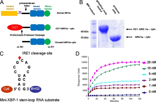
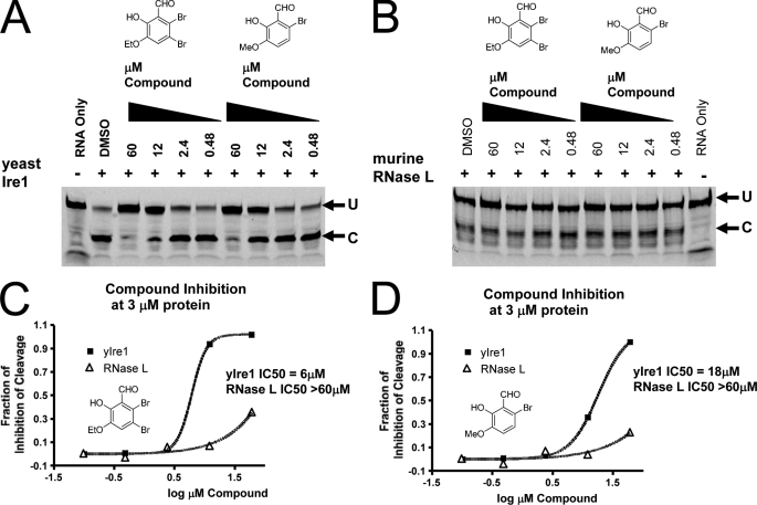
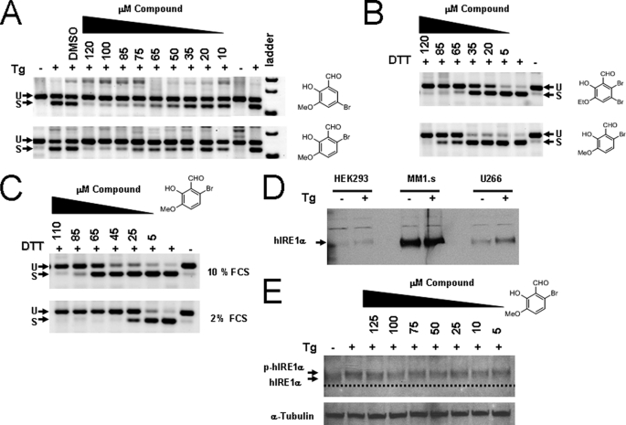
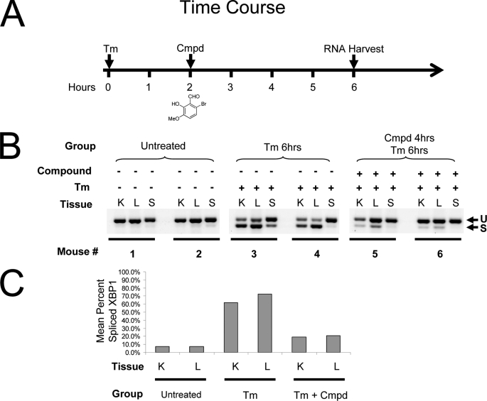
Inositol-requiring enzyme 1 (IRE1) is the most highly conserved signaling node of the unfolded protein response (UPR) and represents a potential therapeutic target for a number of diseases associated with endoplasmic reticulum stress. IRE1 activates the XBP-1 transcription factor by site-specific cleavage of two hairpin loops within its mRNA to facilitate its nonconventional splicing and alternative translation. We screened for inhibitors using a construct containing the unique cytosolic kinase and endoribonuclease domains of human IRE1α (hIRE1α-cyto) and a mini-XBP-1 stem-loop RNA as the substrate. One class compounds was salicylaldehyde analogs from the hydrolyzed product of salicylaldimines in the library. Salicylaldehyde analogs were active in inhibiting the site-specific cleavage of several mini-XBP-1 stem-loop RNAs in a dose-dependent manner. Salicyaldehyde analogs were also active in inhibiting yeast Ire1 but had little activity inhibiting RNase L or the unrelated RNases A and T1. Kinetic analysis revealed that one potent salicylaldehyde analog, 3-ethoxy-5,6-dibromosalicylaldehyde, is a non-competitive inhibitor with respect to the XBP-1 RNA substrate. Surface plasmon resonance studies confirmed this compound bound to IRE1 in a specific, reversible and dose-dependent manner. Salicylaldehydes inhibited XBP-1 splicing induced pharmacologically in human cells. These compounds also blocked transcriptional up-regulation of known XBP-1 targets as well as mRNAs targeted for degradation by IRE1. Finally, the salicylaldehyde analog 3-methoxy-6-bromosalicylaldehyde strongly inhibited XBP-1 splicing in an in vivo model of acute endoplasmic reticulum stress. To our knowledge, salicylaldehyde analogs are the first reported specific IRE1 endoribonuclease inhibitors.


Figures

FIGURE 1.
FIGURE 1.
Recombinant expression and activity of the purified cytosolic human IRE1α kinase endoribonuclease. A, expression strategy for a cytosolic fragment (amino acids 462–977) of human IRE1α (hIRE1α-cyto) using a baculovirus/insect cell system. hIRE1α-cyto was liberated from GST using PreScission protease. B, SDS-polyacrylamide gel electrophoresis analysis of GST-fused and PreScission-liberated hIRE1α-cyto. C, schematic of the mini-XBP-1 stem-loop used as the hIRE1α-cyto substrate for HTS and IC50 analysis; the Cy5 fluorophore was linked to the 5′ end and black hole quencher 2 (BHQ2) was linked to the 3′ end. D, time course analysis of mini-XBP-1 substrate cleavage (100 nm starting concentration) by the indicated concentrations of hIRE1α-cyto.
FIGURE 2.
FIGURE 2.
Active salicylaldimine hit compounds, IC50 curves of their respective salicylaldehydes, and additional analogs used in these studies. Salicylaldimine hit compounds were of the characteristics shown in A, i, where substitutions of X at positions 3 and 5 were di-iodine, di-bromine, or di-chlorine and R was various constituents linked by an imine. ii shows the structure and 50% IC50 profile for a reproduced hit. Performed in triplicate, each compound concentration on the curve is expressed as the mean ± S.D. We determined by LC-MS that the salicylaldimine analogs were hydrolyzing to salicylaldehydes (B) and tested the purified forms without the R groups (C, i–iii). Representative IC50 analysis of salicylaldehyde (C, iv) and a number of analogs (C, v–x) with substituents at positions 3, 5, and 6. PLP (C, xi) and pyridoxal (C, xii), compounds related to salicylaldehydes, were not active.
FIGURE 3.
FIGURE 3.
Cross-reactivity analysis of salicylaldehyde analogs against yeast Ire1 and murine RNase L. Both 3-ethoxy-5,6-dibromosalicylaldehyde and 3-methoxy-6-bromosalicylaldehyde displayed cross-reactivity against yeast Ire1 (A). Yeast Ire1-cyto was preincubated with the indicated concentrations of compound for 1 h at room temperature. Then the 5′ FITC-labeled single hairpin RNA substrate (5′-CAUGUCCGCAGCGCAUG-3′) was added and the reaction incubated for 90 min at room temperature. Reaction mixtures were then resolved by PAGE and fluorescence was visualized by a Typhoon imager. Neither 3-ethoxy-5,6-dibromosalicylaldehyde nor 3-methoxy-6-bromosalicylaldehyde show cross-reactivity against murine RNase L (B). A catalytic fragment (residues 333–651) of murine RNase L expressed and purified from bacteria was preincubated with the indicated concentrations of compound for 1 h at room temperature. Then 5′ FITC-labeled RNase L RNA substrate (5′-C11U2C7-3′) was added and the reaction was incubated for 90 min at room temperature. Reaction mixtures were then resolved by PAGE and fluorescence was visualized by a Typhoon imager. IC50 profiles for 3-ethoxy-5,6-dibromosalicylaldehyde (C) and 3-methoxy-6-bromosalicylaldehyde (D) against yeast Ire1 and RNase L are indicated. Quantification of cleavage was performed by phosphorimager analysis and graphed using GraphPad.
FIGURE 4.
FIGURE 4.
Enzyme kinetics of hIRE1α-cyto, mini-XBP-1 RNA stem-loop, and the inhibitor compound 3-ethoxy-5,6-dibromosalicylaldehyde. Titration of the substrate Cy5-labeled mini-XBP-1 RNA stem-loop as shown in Fig. 1, with constant enzyme (hIRE1α-cyto) concentration and measuring the slope of the initial reaction rates (A, i) determined the Michaelis-Menten constant (Km). Using a Lineweaver-Burk plot (A, ii), the x-axis intercept therefore specified the Km of 0.8 μm for a single mini-XBP-1 RNA stem-loop substrate (Fig. 1C). Titration of both substrate and 3-ethoxy-5,6-dibromosalicylaldehyde demonstrated a non-competitive mode of inhibition relative to the substrate (B). Data in B, i, were plotted as 1/velocity against 1/substrate concentration as a Lineweaver-Burk plot (B, ii). The binding constants Kii and Kis were 71 and 88 nm, respectively. Data are shown from a representative of three separate experiments. Reaction kinetics data were fit using Visual Enzymics software.
FIGURE 5.
FIGURE 5.
Surface plasmon resonance of hIRE1α-cyto, substrate, and compound binding characteristics. Schematic of the XBP-1 RNA stem-loop with an extended stem used for surface plasmon resonance binding experiments is shown (A). Surface plasmon resonance binding profiles with the immobilized reagent on the solid chip surface are indicated at the bottom and the soluble binding partner indicated at the top (A–F). Active hIRE1α-cyto was linked to the Biacore chip by amine coupling and demonstrated specific and dose-dependent binding to the substrate confirming that hIRE1α-cyto was active on the chip (B). When compound 3-ethoxy-5,6-dibromosalicylaldehyde was passed over hIRE1α-cyto on the chip, a specific dose-dependent binding was observed with fast on-fast off kinetics (C). The disassociation constant (Kd) was calculated to be ≈100 nm. Biotinylated stem-loop RNA was immobilized to a streptavidin-coated chip and a large mass change was observed when hIRE1α-cyto was passed over (D). Repeated exposure to soluble hIRE1α-cyto degraded the signal likely due to site-specific cleavage of the stem-loop RNA on the solid surface, therefore, only a single concentration is shown. Passage of 3-ethoxy-5,6-dibromosalicylaldehyde over a chip immobilized with the XBP-1 stem-loop RNA did not give rise to a detectable binding signal (E). When RNase A was coupled to the chip and compound 3-ethoxy-5,6-dibromosalicylaldehyde was passed over the surface, no detectable binding was observed (F).
FIGURE 6.
FIGURE 6.
Inhibition of XBP-1 splicing in human cells using salicylaldehyde analogs. HEK293 cells were left untreated or treated with 300 nm Tg for 3 h. Compounds (right of panel) were added 2 h before Tg at the indicated dose; total RNA was harvested and RT-PCR was performed using human-specific XBP-1 primers flanking the splice site 3 h after stress induction. PCR products were run on 4% agarose gels and stained with ethidium bromide and shown as the inverse image. Spliced (S) and unspliced (U) reaction products of XBP-1 mRNA are designated (A). Human myeloma MM1.s cells were treated with 2 mm DTT as the ER stressing agent and exposed to increasing concentrations of the indicated compound for 2 h (B): both DTT and the compounds were added at the same time. When MM1.s cells were treated in 2% FCS medium as in B, the potency of 3-methoxy-6-bromosalicylaldehyde increased, indicating compounds are partially absorbed to serum proteins (C). Western blot showing relative levels of IRE1α in human cell lines both untreated and treated with 300 nm Tg for 2 h (D). MM1.s myeloma cells had high steady state levels of IRE1α compared with HEK293 and the IgE secreting human myeloma cell line U266. When HEK293 cells were incubated with Tg and increasing concentrations of 3-methoxy-6-bromosalicylaldehyde for 3 h, no change in phosphorylation status was observed (E). The dashed line is a reference to observe the slight decrease in mobility due to phosphorylation (p-hIRE1α). The blot was reprobed with anti-α-tubulin antibody used as a loading control.
FIGURE 7.
FIGURE 7.
In vivo inhibition of XBP-1 splicing by 3-methoxy-6-bromosalicylaldehyde. Time course of in vivo treatment of mice (2 per treatment group) with Tm and 3-methoxy-6-bromosalicylaldehyde (cmpd) (A). CB17 SCID mice were treated with 1 mg/kg of Tm administered by intraperitoneal injection at time 0. Two hours later 3-methoxy-6-bromosalicylaldehyde dissolved in DMSO was delivered by the same route at 50 mg/kg and 4 h later, 6 h from time 0, the kidney (K), liver (L), and spleen (S) were harvested and snap frozen in liquid nitrogen. Tissues were homogenized and total RNA was prepared using TRIzol. RT-PCR was performed using murine-specific primers flanking the XBP-1 mRNA dual hairpin stem-loop and products were separated on a 4% agarose gel. Untreated mice had little or no evidence of XBP-1 splicing (U). Tm treatment induced robust splicing (S) at 6 h and 3-methoxy-6-bromosalicylaldehyde strongly inhibited slicing after 4 h of treatment (B). Scanning and quantification of the bands in B expressed as percent spliced XBP-1 (by dividing intensity of the spliced band by the total intensity of both) shows ∼80% inhibition for the mean of two mice per each group (C).

Similar articles

Cited by

References

    1. Ron D., Walter P. (2007) Nat. Rev. Mol. Cell Biol. 8, 519–529 - PubMed
    1. Lee A. S., Hendershot L. M. (2006) Cancer Biol. Ther. 5, 721–722 - PubMed
    1. Kaufman R. J., Cao S. (2010) EMBO Mol. Med. 2, 189–192 - PMC - PubMed
    1. Hotamisligil G. S. (2010) Cell 140, 900–917 - PMC - PubMed
    1. Cox J. S., Shamu C. E., Walter P. (1993) Cell 73, 1197–1206 - PubMed

Publication types

MeSH terms

LinkOut - more resources