

Lantibiotic transporter requires cooperative functioning of the peptidase domain and the ATP binding domain
- PMID: 21303905
- PMCID: PMC3064170
- DOI: 10.1074/jbc.M110.212704
Lantibiotic transporter requires cooperative functioning of the peptidase domain and the ATP binding domain
Abstract
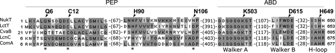
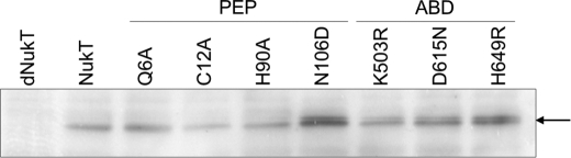
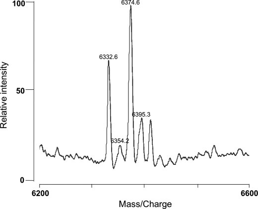
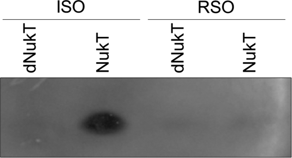
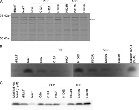
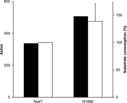
Lantibiotics are ribosomally synthesized and post-translationally modified peptide antibiotics that contain unusual amino acids such as dehydro and lanthionine residues. Nukacin ISK-1 is a class II lantibiotic, whose precursor peptide (NukA) is modified by NukM to form modified NukA. ATP-binding cassette (ABC) transporter NukT is predicted to cleave off the N-terminal leader peptide of modified NukA and secrete the mature peptide. Multiple sequence alignments revealed that NukT has an N-terminal peptidase domain (PEP) and a C-terminal ATP binding domain (ABD). Previously, in vitro reconstitution of NukT has revealed that NukT peptidase activity depends on ATP hydrolysis. Here, we constructed a series of NukT mutants and investigated their transport activity in vivo and peptidase activity in vitro. Most of the mutations of the conserved residues of PEP or ABD resulted in failure of nukacin ISK-1 production and accumulation of modified NukA inside the cells. NukT(N106D) was found to be the only mutant capable of producing nukacin ISK-1. Asn(106) is conserved as Asp in other related ABC transporters. Additionally, an in vitro peptidase assay of NukT mutants demonstrated that PEP is on the cytosolic side and all of the ABD mutants as well as PEP (with the exception of NukT(N106D)) did not have peptidase activity in vitro. Taken together, these observations suggest that the leader peptide is cleaved off inside the cells before peptide secretion; both PEP and ABD are important for NukT peptidase activity, and cooperation between these two domains inside the cells is indispensable for proper functioning of NukT.
Figures
Similar articles
-
ATP-dependent leader peptide cleavage by NukT, a bifunctional ABC transporter, during lantibiotic biosynthesis.J Biosci Bioeng. 2009 Dec;108(6):460-4. doi: 10.1016/j.jbiosc.2009.06.002. J Biosci Bioeng. 2009. PMID: 19914576
-
ATPase activity regulation by leader peptide processing of ABC transporter maturation and secretion protein, NukT, for lantibiotic nukacin ISK-1.Appl Microbiol Biotechnol. 2018 Jan;102(2):763-772. doi: 10.1007/s00253-017-8645-2. Epub 2017 Nov 22. Appl Microbiol Biotechnol. 2018. PMID: 29167920
-
Mapping and identification of the region and secondary structure required for the maturation of the nukacin ISK-1 prepeptide.Peptides. 2009 Aug;30(8):1412-20. doi: 10.1016/j.peptides.2009.05.021. Epub 2009 May 27. Peptides. 2009. PMID: 19481127
-
Type I secretion systems - a story of appendices.Res Microbiol. 2013 Jul-Aug;164(6):596-604. doi: 10.1016/j.resmic.2013.03.011. Epub 2013 Mar 26. Res Microbiol. 2013. PMID: 23541474 Review.
-
Peptide signal molecules and bacteriocins in Gram-negative bacteria: a genome-wide in silico screening for peptides containing a double-glycine leader sequence and their cognate transporters.Peptides. 2004 Sep;25(9):1425-40. doi: 10.1016/j.peptides.2003.10.028. Peptides. 2004. PMID: 15374646 Review.
Cited by
-
Substrate specificity of the lanthipeptide peptidase ElxP and the oxidoreductase ElxO.ACS Chem Biol. 2014 Aug 15;9(8):1718-25. doi: 10.1021/cb5002526. Epub 2014 Jun 6. ACS Chem Biol. 2014. PMID: 24866416 Free PMC article.
-
Structure, Assembly, and Function of Tripartite Efflux and Type 1 Secretion Systems in Gram-Negative Bacteria.Chem Rev. 2021 May 12;121(9):5479-5596. doi: 10.1021/acs.chemrev.1c00055. Epub 2021 Apr 28. Chem Rev. 2021. PMID: 33909410 Free PMC article. Review.
-
One-pot synthesis of class II lanthipeptide bovicin HJ50 via an engineered lanthipeptide synthetase.Sci Rep. 2016 Dec 7;6:38630. doi: 10.1038/srep38630. Sci Rep. 2016. PMID: 27924934 Free PMC article.
-
Genome mining reveals the biosynthetic potential of a novel Lysinibacillus zambalensis sp. nov., isolated from a hyperalkaline spring.Arch Microbiol. 2025 Apr 2;207(5):109. doi: 10.1007/s00203-025-04316-0. Arch Microbiol. 2025. PMID: 40169433 Free PMC article.
-
Ribosomally synthesized and post-translationally modified peptide natural products: new insights into the role of leader and core peptides during biosynthesis.Chemistry. 2013 Jun 10;19(24):7662-77. doi: 10.1002/chem.201300401. Epub 2013 May 10. Chemistry. 2013. PMID: 23666908 Free PMC article.
References
-
- de Vos W. M., Kuipers O. P., van der Meer J. R., Siezen R. J. (1995) Mol. Microbiol. 17, 427–437 - PubMed
-
- Chatterjee C., Paul M., Xie L., van der Donk W. A. (2005) Chem. Rev. 105, 633–684 - PubMed
-
- Nagao J., Asaduzzaman S. M., Aso Y., Okuda K., Nakayama J., Sonomoto K. (2006) J. Biosci. Bioeng. 102, 139–149 - PubMed
-
- Asaduzzaman S. M., Sonomoto K. (2009) J. Biosci. Bioeng. 107, 475–487 - PubMed
-
- Pag U., Sahl H. G. (2002) Curr. Pharm. Des. 8, 815–833 - PubMed
Publication types
MeSH terms
Substances
LinkOut - more resources
Full Text Sources
Other Literature Sources
Research Materials
Miscellaneous
