. 2011 Apr;17(4):697-709.
doi: 10.1261/rna.2509811. Epub 2011 Feb 8.

Understanding ribosome assembly: the structure of in vivo assembled immature 30S subunits revealed by cryo-electron microscopy

Affiliations

Understanding ribosome assembly: the structure of in vivo assembled immature 30S subunits revealed by cryo-electron microscopy

Ahmad Jomaa et al. RNA. 2011 Apr.

Abstract

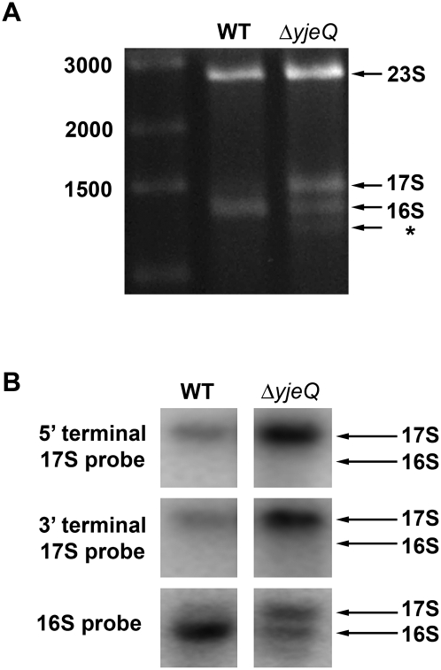
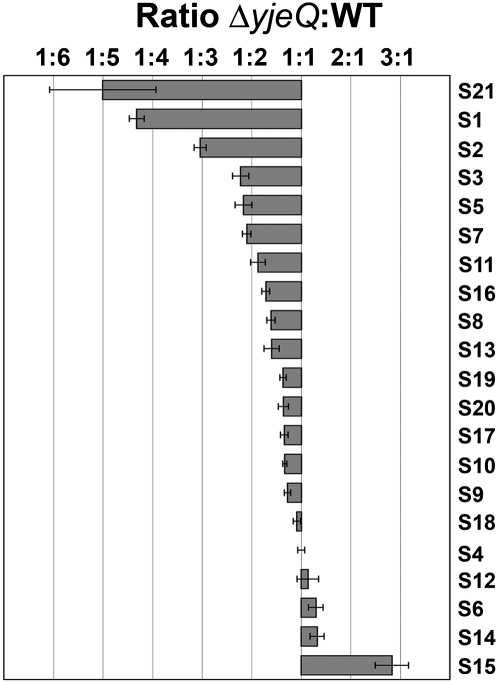
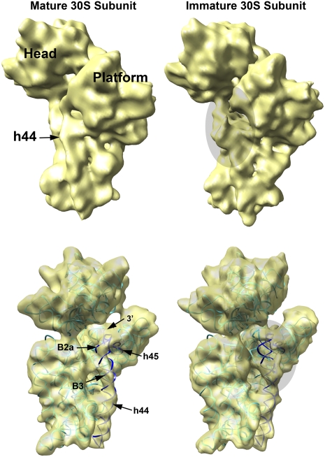
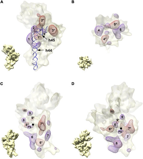
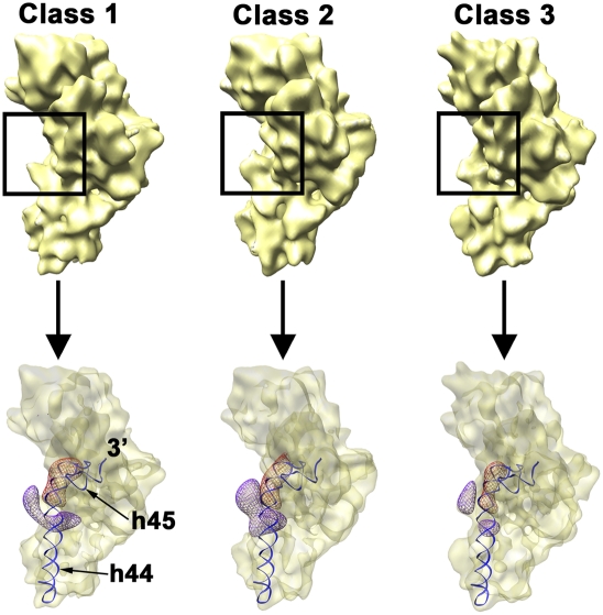
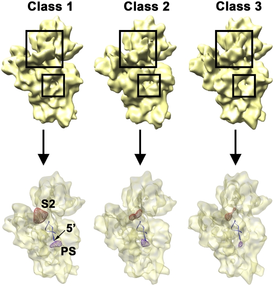
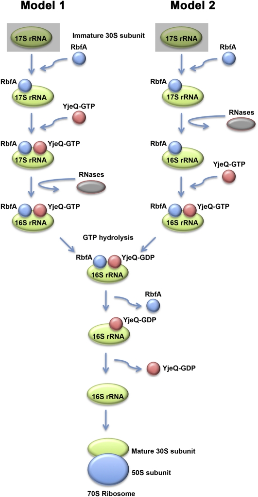
Four decades after early in vitro assembly studies demonstrated that ribosome assembly is a controlled process, our understanding of ribosome assembly is still incomplete. Just as structure determination has been so important to understanding ribosome function, so too will it be critical to sorting out the assembly process. Here, we used a viable deletion in the yjeQ gene, a recognized ribosome assembly factor, to isolate and structurally characterize immature 30S subunits assembled in vivo. These small ribosome subunits contained unprocessed 17S rRNA and lacked some late ribosomal proteins. Cryo-electron microscopy reconstructions revealed that the presence of precursor sequences in the rRNA induces a severe distortion in the 3' minor domain of the subunit involved in the decoding of mRNA and interaction with the large ribosome subunit. These findings suggest that rRNA processing events induce key local conformational changes directing the structure toward the mature assembly. We concluded that rRNA processing, folding, and the entry of tertiary r-proteins are interdependent events in the late stages of 30S subunit assembly. In addition, we demonstrate how studies of emerging assembly factors in ribosome biogenesis can help to elucidate the path of subunit assembly in vivo.


Figures

FIGURE 1.
FIGURE 1.
Characterization of the rRNA from E. coli ΔyjeQ strain. (A) Electrophoretic analysis of rRNA derived from the wild type and yjeQ-null strain cell extracts. The wild-type gel pattern exhibited only two bands with migration corresponding to mature 16S and 23S rRNA. Two additional bands were observed in the yjeQ deletion strain corresponding to a precursor form of 16S rRNA (17S label) and a previously identified species suspected to be a degradation product or an aberrantly processed rRNA (asterisk). The sizes of the fragments of the DNA ladder in the left lane of the gel are indicated. (B) The identity of the precursor 16S rRNA found in the yjeQ-null strain was confirmed by Northern blot analysis using radiolabeled DNA sequence-specific probes directed at the 5′- and 3′-terminal sequences of 17S rRNA. Hybridization of these probes with the rRNA precursor derived from the yjeQ deletion cell extract indicated that the species was indeed unprocessed 17S rRNA with intact 5′ and 3′ terminal sequences. Mobility of the 16S rRNA and 17S rRNA is indicated.
FIGURE 2.
FIGURE 2.
iTRAQ analysis of the 30S subunits purified from ΔyjeQ cells. Small subunit r-proteins. The plot shows the relative levels of the small subunit r-proteins between the 30S subunit purified from ΔyjeQ and wild-type (WT) cells. Relative levels for each protein are expressed as the average ratio ΔyjeQ:WT obtained from two replicas of the experiment. Only peptides that identify proteins with ≥95% confidence were used for the calculation of these ratios. The error bars indicate the standard error of the mean for each ratio.
FIGURE 3.
FIGURE 3.
3D reconstruction of the immature 30S ribosomal subunits purified from ΔyjeQ E. coli cells. Platform view of the cryo-EM maps of the immature (upper right) and mature (upper left) 30S subunits. The immature structure was produced from 42,873 projections of 30S subunits purified from ΔyjeQ cells after 16,180 projections (27.4% of the total) representing mature small subunits were removed from the data set using a supervised classification approach. The mature structure (left) was produced from 19,924 projection images obtained from a homogeneous sample of mature 16S rRNA-containing 30S subunits purified from wild-type cells. Main landmarks of the 30S subunit are indicated. In the panels below, the X-ray structure of the wild-type 30S subunit, shown as a ribbon representation, was fitted into the cryo-EM maps of both the mature (left) and immature (right) 30S subunits to illustrate their divergences. In the X-ray structure, the rRNA is shown in cyan except helices 44 and 45, which are dark blue. The r-proteins are not shown. Main landmarks of the 30S subunit indicated include helix 44 (h44), helix 45 (h45), the intersubunit bridges B2a (B2a) and B3 (B3) important for the interaction with the 50S subunit, and the 3′ terminus of the 16S rRNA molecule (3′). The shadowed ovals in the right panels show the area in the helix 44 and decoding center distorted in the EM map of the immature 30S subunit.
FIGURE 4.
FIGURE 4.
Difference map analysis of the immature 30S subunit cryo-EM structure. Overlay of the forward and reverse difference maps. Densities from the forward (violet mesh) and reverse (red mesh) difference maps are shown contained in a semitransparent surface representation of the immature 30S subunit structure from ΔyjeQ cells. Matching densities are identified with the same number, and the density in the reverse map is identified by an asterisk (*). (A) The densities present in the 3′ minor (helices 44 and 45) and central domains of the two difference maps. Similarly, panel B displays only the densities present in the 3′ major domain of these difference maps, and panels C and D show two views of the densities existing on the 5′ domain. The small cryo-EM map in the bottom left corner of each panel is an aid for orientation and represents the immature 30S subunit in the same view as the difference maps in the corresponding panel. Helices 44 (h44) and 45 (h45) are also shown as a ribbon representation in A to illustrate the displacement of helix 44 in the immature 30S subunit structure. The 5′ end of the mature 16S rRNA is indicated with a black dot in C and D. The density labeled as “S2” in C and D indicates the amount of density corresponding to this protein missing in the immature structure. The density labeled as “PS” in the same panels represents the additional density existing in the immature structure due to the presence of the 5′ precursor sequence of the rRNA.
FIGURE 5.
FIGURE 5.
Conformational variability of the immature 30S subunit from ΔyjeQ cells. Platform view of the three cryo-EM structures (labeled from class 1 to class 3) representing three subpopulations of immature 30S subunits present in ΔyjeQ cells (top). These 3D reconstructions were produced from 13,447 (class 1), 12,570 (class 2), and 11,931 (class 3) projections and refined to 14.1 Å (class 1), 15.4 Å (class 2), and 14.7 Å (class 3) resolution. The framed region in each map highlights the area of helix 44 that is distorted in the three subpopulations. The bottom panel illustrates the degree of distortion of helix 44 in each cryo-EM map. The density corresponding to the upper segment of helix 44 in each conformational state is shown as a violet mesh. The displacement of helix 44 in each conformation is shown by comparison to the position of the helix 44 in the mature 30S subunit shown as a red mesh. Helices 44 (h44) and 45 (h45) in the mature structure are also shown for reference.
FIGURE 6.
FIGURE 6.
Variability of the density representing the S2 r-protein and 5′ precursor sequences in the immature 30S subunit from ΔyjeQ cells. Back view of the three cryo-EM structures (labeled from class 1 to class 3) representing three subpopulations of immature 30S subunits present in ΔyjeQ cells (top). The framed regions highlight the location of the S2 r-protein and the 5′ mature end of the 16S rRNA. The dissimilar amount of density in these two locations in the cryo-EM maps is shown in the bottom panel. The density corresponding to the S2 r-protein (red mesh) from the reverse difference map (i.e., mature 30S subunit map minus immature 30S map) and the one corresponding to the 5′ precursor sequence (violet mesh) from the forward difference map (i.e., immature 30S subunit map minus mature 30S map) for each cryo-EM map are displayed within a semitransparent surface of the 30S subunit structure. The first one indicates the amount of density corresponding to the S2 r-protein missing in each immature structure. Conversely, the second one shows the additional density existing in each structure due to the presence of the 5′ precursor sequence. The first 20 nucleotides of the 5′ end of the mature 16S rRNA are shown as a ribbon representation.
FIGURE 7.
FIGURE 7.
Placement of the structurally characterized immature 30S subunits in the current biogenesis model of the 30S subunit. The diagram describes the likely order of events involving YjeQ in the late stages of assembly of the 30S subunit and processing of the 17S rRNA. A recent publication (Goto et al. 2011) has suggested that YjeQ mediates this process in conjunction with RbfA, and thus, this protein is included in the diagram. RbfA seems to bind first to an immature form of the 30S subunit facilitating the subsequent entry of YjeQ-GTP to the maturing subunit. At this moment, there is not enough experimental evidence yet to establish whether the processing of the 17S rRNA occurs before (model 2) or after (model 1) YjeQ binding to the immature 30S subunit. The cryo-EM reconstruction of the immature 30S subunit purified from the ΔYjeQ strain that is presented constitutes the structure of a 17S rRNA–containing 30S subunit prior to rRNA processing and could possibly represent the structure of the immature 30S subunit bound by RbfA (shadowed squares in models 1 and 2).

Similar articles

Cited by

References

    1. Adilakshmi T, Bellur DL, Woodson SA 2008. Concurrent nucleation of 16S folding and induced fit in 30S ribosome assembly. Nature 455: 1268–1272 - PMC - PubMed
    1. Aebi U, Pollard TD 1987. A glow discharge unit to render electron microscope grids and other surfaces hydrophilic. J Electron Microsc Tech 7: 29–33 - PubMed
    1. Ban N, Nissen P, Hansen J, Moore PB, Steitz TA 2000. The complete atomic structure of the large ribosomal subunit at 2.4 Å resolution. Science 289: 905–920 - PubMed
    1. Borovinskaya MA, Pai RD, Zhang W, Schuwirth BS, Holton JM, Hirokawa G, Kaji H, Kaji A, Cate JH 2007. Structural basis for aminoglycoside inhibition of bacterial ribosome recycling. Nat Struct Mol Biol 14: 727–732 - PubMed
    1. Brown ED 2005. Conserved P-loop GTPases of unknown function in bacteria: an emerging and vital ensemble in bacterial physiology. Biochem Cell Biol 83: 738–746 - PubMed

Publication types

MeSH terms

LinkOut - more resources