. 2011 Apr;152(4):1434-47.
doi: 10.1210/en.2010-1368. Epub 2011 Feb 8.

GPR30 activation opposes estrogen-dependent uterine growth via inhibition of stromal ERK1/2 and estrogen receptor alpha (ERα) phosphorylation signals

Affiliations

GPR30 activation opposes estrogen-dependent uterine growth via inhibition of stromal ERK1/2 and estrogen receptor alpha (ERα) phosphorylation signals

Fei Gao et al. Endocrinology. 2011 Apr.

Abstract

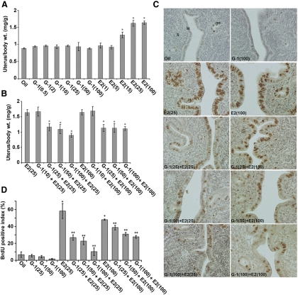
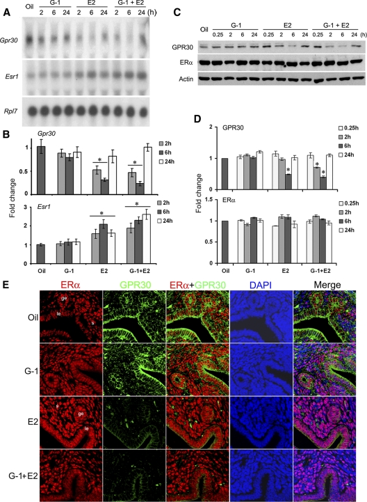
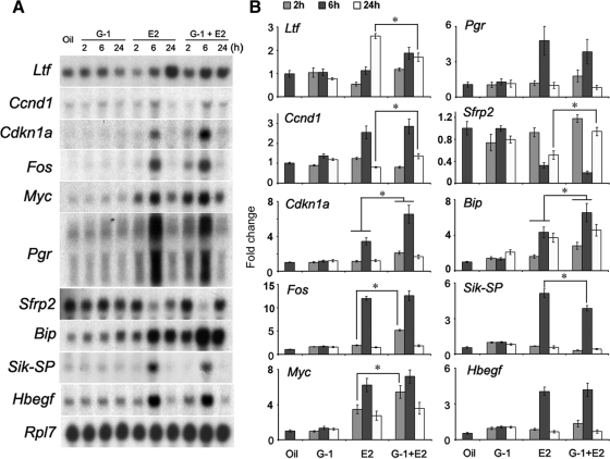
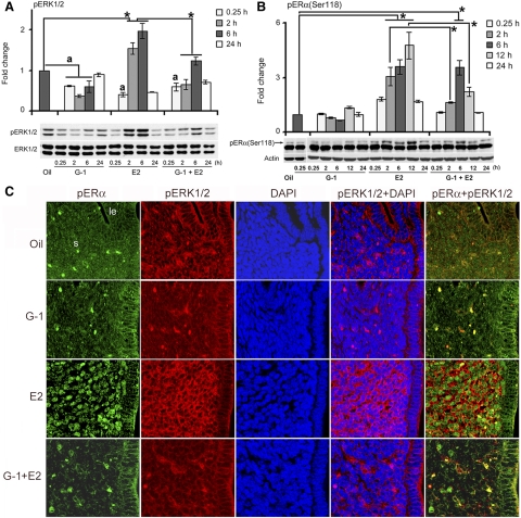
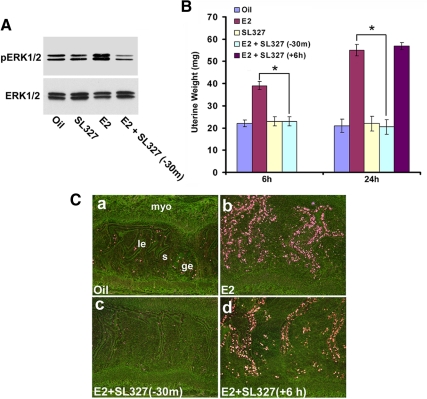
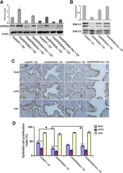
Although estradiol-17β (E2)-regulated early and late phase uterine responses have been well defined, the molecular mechanisms linking the phases remain poorly understood. We have previously shown that E2-regulated early signals mediate cross talk with estrogen receptor (ER)-α to elicit uterine late growth responses. G protein-coupled receptor (GPR30) has been implicated in early nongenomic signaling mediated by E2, although its role in E2-dependent uterine biology is unclear. Using selective activation of GPR30 by G-1, we show here a new function of GPR30 in regulating early signaling events, including the inhibition of ERK1/2 and ERα (Ser118) phosphorylation signals and perturbation of growth regulation under the direction of E2 in the mouse uterus. We observed that GPR30 primarily localizes in the uterine epithelial cells, and its activation alters gene expression and mediates inhibition of ERK1/2 and ERα (Ser118) phosphorylation signals in the stromal compartment, suggesting a paracrine signaling is involved. Importantly, viral-driven manipulation of GPR30 or pharmacological inhibition of ERK1/2 activation effectively alters E2-dependent uterine growth responses. Overall, GPR30 is a negative regulator of ERα-dependent uterine growth in response to E2. Our work has uncovered a novel GPR30-regulated inhibitory event, which may be physiologically relevant in both normal and pathological situations to negatively balance ERα-dependent uterine growth regulatory functions induced by E2.


Figures

Fig. 1.
Fig. 1.
G-1 by itself is not stimulatory but can antagonize E2-induced uterine growth responses. Dosage-dependent studies were conducted in adult ovariectomized mice after sc injections of oil (vehicle control), G-1 (0.5, 2, 10, 25, 50, or 100 μg/mouse), E2 (1, 5, 15, 25, or 100 ng/mouse) (A), or G-1 (10, 25, 50, or 100 μg/mouse) 30 min before an injection of E2 (25 ng/mouse) or E2 (100 ng/mouse) (B). Mice were killed 24 h after the last injection. Uterine wet weights from five to 10 mice for each group were analyzed. The error bars represent ses. *, Values are statistically different compared with the vehicle control (P < 0.01, ANOVA followed by Newman-Keul's multiple range test). C, G-1 inhibits E2-induced uterine epithelial cell proliferation. Adult ovariectomized mice were injected with different agents as described above, and BrdU was injected 2 h before the animals were killed. Formaldehyde-fixed paraffin-embedded tissue sections were stained for BrdU incorporation as described in Materials and Methods. Representative tissue sections are shown and reddish brown nuclear deposits indicate the sites of positive immunostaining. Pictures were taken at ×200. le, Luminal epithelium; ge, glandular epithelium; s, stroma cells. D, Quantitation of BrdU-positive cells. The percentage of BrdU labeling index was determined for the luminal and glandular epithelial cells after counting at least 500 cells per animal on consecutive fields. The data presented after the analysis of at least five to 10 mice for each group. The error bars represent ses. *, Values are statistically different (P < 0.01) against the oil (control). **, Values are statistically different (P < 0.05) against E2 (25 ng/mouse or 100 ng/mouse) based on ANOVA followed by Newman-Keul's multiple range test.
Fig. 2.
Fig. 2.
Uterine expression of GPR30 is suppressed by E2 treatment. Adult ovariectomized mice were given a single injection of G-1 (100 μg/mouse), E2 (25 ng/mouse), or the same doses of G-1 plus E2 and killed at the indicated times. Mice injected with oil and killed after 6 h served as vehicle controls. Temporal effects of uterine mRNA (A) and protein (C) levels for the expression of Esr1 and Gpr30 were analyzed by Northern and Western blotting techniques, respectively. These experiments were repeated four times with independent samples, and a representative blot is presented. As previously reported by us (9, 10), the quantitation of mRNA transcripts was achieved by direct analysis of radioactivity bound to the hybridized bands using a radioimage analysis system (Ambis Systems, San Diego, CA). The quantitation of protein levels from the Western blots was determined by densitometric scanning. Fold changes in mRNA (B) or protein (D) levels were calculated against oil and normalized by Rpl7 or actin levels, respectively. The error bars represent ses. *, Values are statistically different (P < 0.05, ANOVA followed by Newman-Keul's multiple range test) against the oil-treated group. E, GPR30 is colocalized with ERα in uterine epithelial cells. Adult ovariectomized mice were treated as above and killed after 6 h. Formalin-fixed, paraffin-embedded uterine sections were incubated with primary antibodies for ERα and GPR30 followed by incubation of secondary antibodies tagged with Texas-red (red) and Cy2 (green), respectively. Nuclei were stained by 4′,6′-diamidino-2-phenylindole (DAPI) (blue). Merged images are shown with overlapping colors. No immunostaining was noted when sections were incubated with preimmune serum instead of primary antibody (data not shown). Pictures were taken at ×200. le, Luminal epithelium; ge, glandular epithelium; s, stroma cells. These experiments were repeated at least three times with similar results.
Fig. 3.
Fig. 3.
Activation of GPR30 modulates E2-regulated uterine gene expression. Adult ovariectomized mice were treated as in Fig. 2 and killed at the indicated times. Mice injected with oil and killed after 6 h served as vehicle controls. A, Northern blot hybridization for the analysis of temporal effects of the above agents on uterine mRNA expression of Ltf, cdkn1a, Ccnd1, Fos, Myc, Pgr, Sfrp2, Bip, Sik-SP, and Hbegf genes. Rpl7 was used as an internal control gene. These experiments were repeated four times with independent RNA samples, and a representative blot is presented. B, Fold changes in mRNA levels were determined as described in Fig. 2. The error bars represent ses. *, Values are statistically different (P < 0.05, ANOVA followed by Newman-Keul's multiple range test) between the compared groups.
Fig. 4.
Fig. 4.
G-1 mediated activation of GPR30 inhibits E2-induced uterine ERK and ERα phosphorylation in the stromal compartment. Adult ovariectomized mice were treated as in Fig. 2 and killed at the indicated times. Mice injected with oil and killed after 6 h served as vehicle controls. Western blot analysis of whole uterine tissue extracts for pERK1/2 (A) and pERα (Ser118) (B). These experiments were repeated four times with independent samples, and a representative blot is presented. Fold changes in the levels of pERK1/2 and pERα (Ser118) were compared against the control, and the values were determined after normalization with ERK1/2 and actin levels, respectively. The error bars represent ses. *, Values are statistically different (P < 0.05, ANOVA followed by Newman-Keul's multiple range test) between the compared groups. a, Values are significantly different (P < 0.05, ANOVA followed by Newman-Keul's multiple range test) compared with oil treated group. C, Immunofluorescence analyses of pERK1/2 and pERα (Ser118) in uteri of ovariectomized mice treated with agents as described in Fig. 2 and analyzed at 2, 6, and 12 h after the last injection. Formalin-fixed, paraffin-embedded uterine sections were incubated with primary antibodies for pERK1/2 and pERα (Ser118) followed by incubation of secondary antibodies tagged with Cy3 (red) and Cy2 (green), respectively. Nuclei were stained with 4′,6′-diamidino-2-phenylindole (DAPI) (blue). Merged images are shown with overlapping colors. Representative pictures are shown for the 2-h-treated groups. Similar results were obtained for the 6- and 12-h-treated groups (data not shown). No immunostaining was noted when sections were incubated with preimmune serum instead of primary antibody (data not shown). Pictures were taken at ×200. le, Luminal epithelium; ge, glandular epithelium; s, stroma cells. These experiments were repeated at least three times with similar results.
Fig. 5.
Fig. 5.
Pharmacological inhibition of ERK activation abrogates E2-dependent uterine responses. Adult ovariectomized mice were given injections of oil, E2 (100 ng/mouse), SL327 (160 mg/kg), or SL327 (160 mg/kg) 30 min before (−30 m) or 6 h after (+6 h) E2 (100 ng/mouse). A, Western blotting (at 6 h time point) confirms the appropriate inhibition of E2-dependent uterine pERK1/2 activation by SL327. B, Uterine wet weights were recorded at indicated times after the last injection. Uterine wet weights from five to seven mice for each group were analyzed. *, Values are statistically different between the compared groups (P < 0.05) based on ANOVA followed by Newman-Keul's multiple range test. C, Autoradiographic analysis of uterine cell proliferation by 3H-thymidine incorporation. Dark-field photomicrographs of representative tissue sections are shown. Pictures were taken at ×100. le, Luminal epithelium; ge, glandular epithelium; s, stroma; and myo, myometrium. These experiments were repeated at least three times with similar results.
Fig. 6.
Fig. 6.
Adenoviral vector-driven manipulation of GPR30 causes alteration of ERK activation and uterine epithelial cell proliferation induced by E2. A, Ovariectomized mice were administrated with intraluminal injections of adenoviruses empty vector (rAdGFP; control), rAdGPR30(S), AdGPR30(AS), or rAdGPR30(S+AS), and uterine tissues were collected after injections of oil or E2 (25 ng/mouse) at 6 and 24 h. This experiment used at least five mice for each group and repeated four times. Western blotting (at 6 h time point) confirms the appropriate regulation of uterine expression for GPR30 levels (A) and E2-dependent regulation of ERK1/2 activation (B). Fold changes of protein levels were determined by densitometric scanning and calculated against the control virus, and the values were normalized by either actin or ERK1/2 levels, respectively. C, Analysis of uterine epithelial cell proliferation (at 24 h time point) was examined by immunostaining with BrdU, pHH3, and Ki67. Representative tissue sections are shown, and reddish brown nuclear deposits indicate the sites of positive immunostaining. Pictures were taken at ×200. le, Luminal epithelium; ge, glandular epithelium; s, stroma cells. D, Quantitation of BrdU-, pHH3-, and Ki67-positive cells was determined for the luminal and glandular epithelial cells after counting at least 500 cells per animal on consecutive fields. The data are presented after the analysis of at least five to 10 mice for each group. The error bars represent ses. *, Values are statistically different (P < 0.05) between compared groups, based on ANOVA followed by Newman-Keul's multiple range test.

Similar articles

Cited by

References

    1. Couse JF, Korach KS. 1999. Estrogen receptor null mice: what have we learned and where will they lead us? Endocr Rev 20:358–417 - PubMed
    1. Stampfer MJ, Colditz GA, Willett WC, Manson JE, Rosner B, Speizer FE, Hennekens CH. 1991. Postmenopausal estrogen therapy and cardiovascular disease. Ten-year follow-up from the Nurses' Health Study. N Engl J Med 325:756–762 - PubMed
    1. McDonnell DP, Norris JD. 2002. Connections and regulation of the human estrogen receptor. Science 296:1642–1644 - PubMed
    1. Tsai MJ, O'Malley BW. 1994. Molecular mechanisms of action of steroid/thyroid receptor superfamily members. Annu Rev Biochem 63:451–486 - PubMed
    1. Beato M, Herrlich P, Schütz G. 1995. Steroid hormone receptors: many actors in search of a plot. Cell 83:851–857 - PubMed

Publication types

MeSH terms

Substances