. 2011 Apr;152(4):1640-51.
doi: 10.1210/en.2010-1121. Epub 2011 Feb 8.

Essential roles of androgen signaling in Wolffian duct stabilization and epididymal cell differentiation

Affiliations

Essential roles of androgen signaling in Wolffian duct stabilization and epididymal cell differentiation

Aki Murashima et al. Endocrinology. 2011 Apr.

Abstract

The epididymis is a male accessory organ and functions for sperm maturation and storage under the control of androgen. The development of the epididymis is also androgen dependent. The Wolffian duct (WD), anlagen of the epididymis, is formed in both male and female embryos; however, it is stabilized only in male embryos by testicular androgen. Androgen drives subsequent differentiation of the WD into the epididymis. Although the essential roles of androgen in WD masculinization and epididymal function have been established, little is known about cellular events regulated precisely by androgen signaling during these processes. It is also unclear whether androgen signaling, especially in the epithelia, has further function for epididymal epithelial cell differentiation. In this study we examined the cellular death and proliferation controlled by androgen signaling via the androgen receptor (AR) in WD stabilization. Analyses using AR knockout mice revealed that androgen signaling inhibits epithelial cell death in this process. Analysis of AP2α-Cre;AR(flox/Y) mice, in which AR function is deleted in the WD epithelium, revealed that epithelial AR is not required for the WD stabilization but is required for epithelial cell differentiation in the epididymis. Specifically, loss of epithelial AR significantly reduced expression of p63 that is essential for differentiation of basal cells in the epididymal epithelium. We also interrogated the possibility of regulation of the p63 gene (Trp63) by AR in vitro and found that p63 is a likely direct target of AR regulation.


Figures

Fig. 1.
Fig. 1.
Spatiotemporal expression of AR during murine male and female WD development. A–H, Immunostaining (brown signal, DAB) for AR in sectioned male embryos from E12.5 to E18.5. The WD epithelia are encircled with a black dashed line and are surrounded by mesenchyme. Representative sections from cranial and caudal regions of the developing duct are shown for each embryonic stage. I–L, Immunostaining of AR in female embryos at E12.5 and E14.5. AR expression was also observed in the cranial region of WD mesenchyme as the case of male embryos at E12.5 and E14.5. M, Male cranial WD for negative control sample without anti-AR antibody (E12.5). Scale bar, 50 μm (A–M). N, The structure of the male WD at E14.5. Cranial and caudal regions are indicated (WD epithelium was stained for Wnt9b expression by in situ hybridization). t, Testis.
Fig. 2.
Fig. 2.
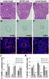
AR KO male embryos show WD regression with prominent epithelial apoptosis similar to the control females. A–C, Histological sections of the cranial region of the WD at E14.5 in control male (AR+/Y), control female (AR+/− or AR+/+), and AR KO (AR−/Y) specimens. D–F, Apoptosis (brown signal, DAB) in the WD assayed by TUNEL at E14.5. G–I, Proliferating cells in WD were detected by BrdU incorporation at E14.5. Scale bar, 100 μm (A–I). Dashed lines, WD; nuclear staining, Hoechst 33342. J, Quantitative assessment of TUNEL-positive cells in different regions of the WD epithelia at E14.5. K, Quantitative assessment of BrdU-positive cells in different regions of the WD epithelia at E14.5. Four embryonic specimens were analyzed using three to four serial sections in each region. (means ± se). *, P < 0.05; **, P < 0.01; ***, P < 0.001.
Fig. 3.
Fig. 3.
AR mutation in the WD epithelium does not affect WD stabilization and WD differentiation during embryogenesis. A–D, Immunohistochemical detection of AR in the cranial WD and epididymis at E14.5 and E18.5. In AP2α-Cre;ARflox/Y embryos, AR expression was undetectable in the WD epithelium. E and F, Gross anatomical structures of the control and AP2α-Cre;ARflox/Y epididymides at E18.5. Arrowheads, Epididymis; t, testis. G and H, Quantitative assessment of TUNEL- and BrdU-positive cells in the WD epithelia at E14.5. Three embryonic specimens were analyzed using three to four serial sections in each region. I and J, Immunofluorescent detection of smooth muscle actin (SMA; green) and smooth muscle myosin (SMM; red) in the caput epididymis at E18.5. Nuclei were stained with Hoechst 33342. epi, Epithelium; mes, mesenchyme. Scale bars, 100 μm (A–D, I, and J).
Fig. 4.
Fig. 4.
Epithelial cell differentiation is impaired in the AP2α-Cre;ARflox/Y adult epididymides. A and B, Gross anatomy of the epididymides of AP2α-Cre;ARflox/Y and control males at P35. Arrows, Epididymis; cap, caput epididymis; cau, cauda epididymis; t, testis. C and D, Toluidine blue staining of transverse sections in caput epididymides at P35 (black arrowheads, narrow cells; yellow arrowheads, basal cells). The boxed areas indicate high-magnification images. epi, Epithelium; mes, mesenchyme; L, lumen; N, narrow cell; P, principal cell; yellow arrow, basal cell. Scale bars, 10 μm. Immunohistochemistry for differentiation markers was performed in the caput (E–L) and cauda (E′–L′) epididymides at P35. AQP9 labels principal cells in the control (E and E′). Its expression was decreased in the mutant (F and F′). K14 and p63 were expressed in basal cells of control epididymides (G, G′, I, and I′). Their expressions were reduced in caput but not in cauda of mutant epididymides (H, H′, J, and J′). Foxi1 expression was observed in both control and mutant epithelia (K, L, K′, and L′). *, Lumen. Scale bars, 200 μm (E–L and E′–L′).
Fig. 5.
Fig. 5.
p63 KO epididymides display abnormal basal cell differentiation. A and B, Histological sections of epididymides 1 month after transplant. Boxed areas are magnified images. Morphologically defined basal cells (A, arrowhead) were not observed in the caput region of the p63 KO epididymides (B). Elongated/flat nuclei of mesenchymal cells were distinct from those of basal cells. C–H, Immunohistochemistry for epithelial cell differentiation markers, K14 (for basal cell, C and D), AQP9 (for principal cell, E and F), and Foxi1 (for narrow and clear cells, G and H). epi, Epithelium; mes, mesenchyme; L, lumen. Scale bars (A and B), 10 μm (A and B); 200 μm (C–H).
Fig. 6.
Fig. 6.
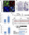
Possible AR functions for the expression of p63 in the epididymis. A, Immunofluorescent staining for AR (green), p63 (red), and nuclei (Hoechst 33342, blue) in CAG-CreERTM;ARflox/Y mutant epididymis at P15. TM injection at E13.5 results in mosaic AR expression in the ductal epithelium. AR− cells were not p63+ (arrowheads; see also Supplemental Fig. 2B). Almost all p63+ cells are also expressed AR. Epi, Epithelium; mes, mesenchyme; L, lumen. B, Immunohistochemical detection of p63 in the developing epididymis of ICR mice. p63 was barely detectable at E18.5 but was present throughout the epididymis at P5. Dashed line, Ductal tube. C, HaCaT cells were transfected with the mouse AR expression vector and treated with 10−8 m DHT or vehicle (dimethylsulfoxide). Expression levels of ΔNp63 were increased after DHT treatment (means ± se). *, P < 0.05. D, Diagram of the Trp63 locus. The PARR is indicated. TAp63 transcripts start from exon 1, whereas ΔNp63 transcripts start from the alternative exon 3′ (arrows). A VISTA plot of the alignment of Trp63 intron 3 of several species is shown. PARR is indicated by gray bar. Exons are indicated in blue, and the conserved noncoding regions are in pink. The mouse genomic sequence (m) was compared with the human (h), rat (r), chicken (c), and dog (d) sequences by MultiLagan (http://lagan.stanford.edu/lagan_web/index.shtml) and is represented on the x-axis. The y-axis represents percent identity, with a scale between 50 and 100%. E, PARR was cloned into TK promoter/luciferase expression constructs. HaCaT cells were transfected with the reporter constructs and mouse AR expression vector. PARR element activated the expression of a luciferase reporter in response to androgen signaling (means ± se). *, P < 0.001. Luciferase activity was not augmented by DHT treatment when AR expression vector were omitted. F, Schematic representation of PARR reporter construct. Mutations were introduced into putative AREs in the PARR (indicated as ARE1 and ARE2, respectively). Fold activation of luciferase activities by 10−8 m DHT treatment was compared between PARR and PARRs harboring the mutation (means ± se). *, P < 0.005.

Similar articles

Cited by

References

    1. Jones RC. 2002. Evolution of the vertebrate epididymis. In: Robaire B, Hinton BT. ed. The epididymis. New York: Kluwer Academic/Plenum Publishers; 11–33
    1. Hinton BT, Palladino MA. 1995. Epididymal epithelium: its contribution to the formation of a luminal fluid microenvironment. Microsc Res Tech 30:67–81 - PubMed
    1. Zhou CX, Zhang YL, Xiao L, Zheng M, Leung KM, Chan MY, Lo PS, Tsang LL, Wong HY, Ho LS, Chung YW, Chan HC. 2004. An epididymis-specific β-defensin is important for the initiation of sperm maturation. Nat Cell Biol 6:458–464 - PubMed
    1. Staack A, Donjacour AA, Brody J, Cunha GR, Carroll P. 2003. Mouse urogenital development: a practical approach. Differentiation 71:402–413 - PubMed
    1. Jost A. 1970. Hormonal factors in the sex differentiation of the mammalian foetus. Philos Trans R Soc Lond B Biol Sci 259:119–130 - PubMed

Publication types

MeSH terms