. 2011 Feb 15;71(4):1282-91.
doi: 10.1158/0008-5472.CAN-10-2480. Epub 2011 Feb 8.

PIPKIγ regulates β-catenin transcriptional activity downstream of growth factor receptor signaling

Affiliations

PIPKIγ regulates β-catenin transcriptional activity downstream of growth factor receptor signaling

Mark Schramp et al. Cancer Res. .

Abstract

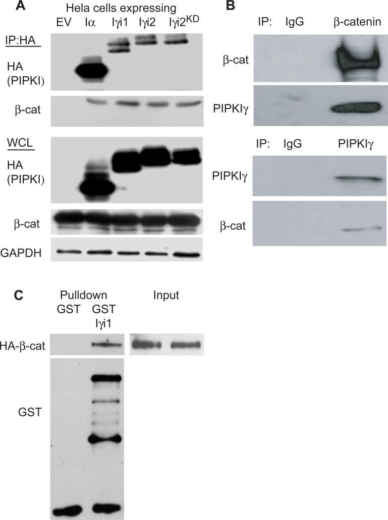
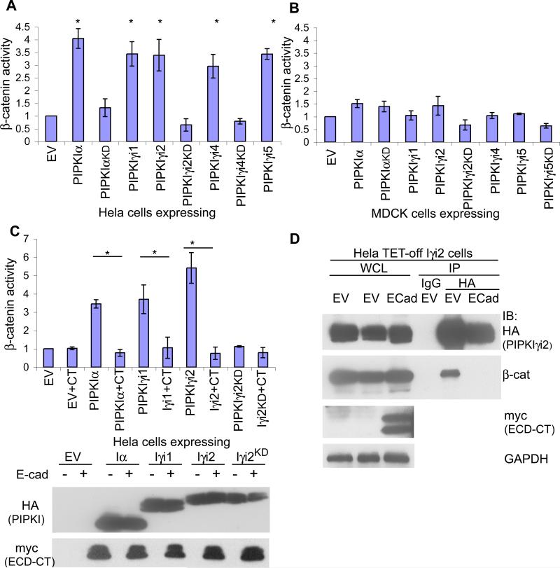
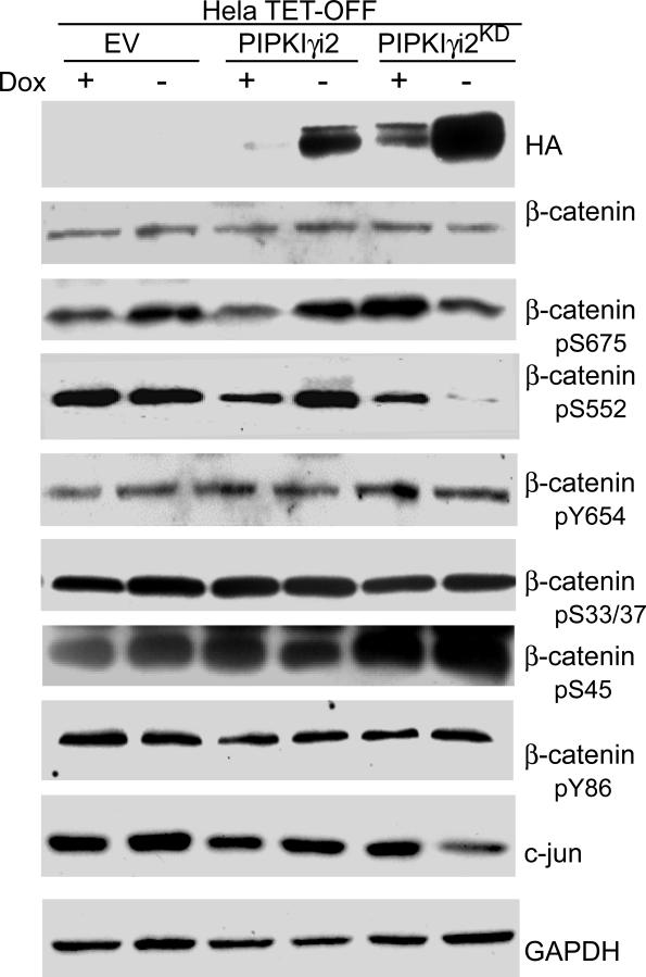
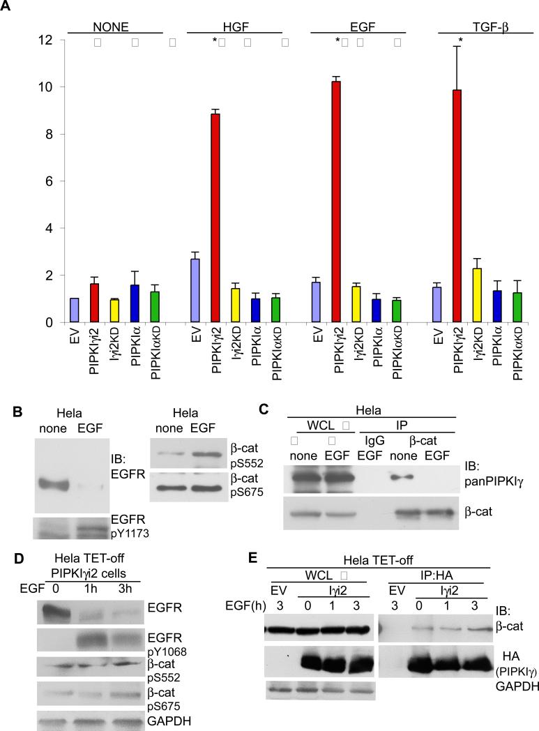
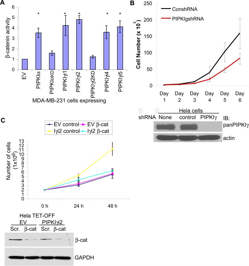
Increased β-catenin transcriptional activity downstream of the Wnt/Wingless signaling pathway has been observed in many human tumors, most notably colorectal carcinomas. However, β-catenin activation is also observed in many human malignancies with no observable Wnt activity. Wnt-independent pathways that activate β-catenin remain undefined, yet have the potential to play a significant role during tumorigenesis. Here, we report that phosphotidylinositol phosphate kinase Iγ (PIPKIγ), an enzyme that generates phosphoinositide messengers in vivo, directly associates with β-catenin and increases β-catenin activity downstream of growth factor stimulation. PIPKIγ expression and kinase activity enhance β-catenin phosphorylation on residues that promote nuclear importation and transcriptional activity. Lastly, we show that β-catenin is required for PIPKIγ-dependent increased cell proliferation. These results reveal a novel mechanism in which PIPKIγ expression and catalytic activity enhance β-catenin nuclear translocation and expression of its target genes to promote tumorigenic phenotypes.


Figures

Figure 1
Figure 1. PIPKIγ directly associates with β–catenin independent of E-cadherin
(A) Hela cells were transiently transfected with empty vector (EV) or the HA-tagged PIPKI shown and an interaction with β-catenin assayed by immunoprecipitation (IP) using anti-HA antibody. Whole cell lysates (WCL) were probed with anti-HA, anti-β-catenin and anti-GAPDH to monitor transfection and protein levels. (B) Endogenous β-catenin and PIPKIγ_i1 were co-IP’ed from A431D cells. (C) Recombinant GST or GST-β-catenin was assayed for interaction with purified PIPKIγ_i1 by in vitro pulldown assays.
Figure 2
Figure 2. PIPKIγ expression stimulates β-catenin transcriptional activity in mesenchymal-like cells lacking E-cadherin
β-catenin transcriptional activity was measured in (A) Hela or (B) MDCK cells transiently transfected with either empty vector (EV) or the HA-tagged PIPKI shown (* represents p-value ≤ 0.001). (C) Hela cells were treated as in (A) however either empty vector (EV) or E-cadherin cytoplasmic domain (CT) included in the transfection where indicated (* represents pvalue ≤ 0.01). Representive western blots show the expression of PIPKIs with myc-tagged E-cadherin C-terminus. (D) Hela TET-off cells expressing PIPKIγ_i2 were transfected with either empty vector or myc-E-cadherinCT where indicated. Lysates of equal protein concentration were IP’d using anti-HA to determine how the interaction between PIPKIγ_i2 and β-catenin was affected upon expression of E-cadherin. For all graphs n≥3, error bars=std dev.
Figure 3
Figure 3. PIPKIγ_i2 generation of phosphoinositides increases the nuclear accumulation of β-catenin
Hela cells grown on glass coverslips for 24 h were transiently transfected with empty vector (EV) or HA-tagged PIPKIγ_i2 or PIPKIγ_i2KD where indicated. 24 h after transfection, cells were fixed and stained with anti-HA to monitor PIPKIγ expressing cells, anti-β-catenin and DAPI (pseudocolored red). All images were taken with a 60X objective.
Figure 4
Figure 4. PIPKIγ expression stimulates the phosphorylation of β-catenin at sites known to initiate nuclear translocation and transcriptional activation
Hela TETOFF cells stably expressing empty vector (EV) or HA-tagged PIPKIγ_i2 or PIPKIγ_i2KD were maintained in DMEM+10%FBS+doxycycline. To initiate PIPKIγ_i2 expression, media was replaced with fresh media +/- dox. where indicated. 24 h later, whole cells lysates were prepared and probed with the antibodies mentioned.
Figure 5
Figure 5. PIPKIγ activates β-catenin upon growth factor stimulation
(A) β-catenin transcriptional activity was measured in Hela cells transiently transfected with empty vector (EV) or the HA-tagged PIPKI shown. 24 h after transfection, cells were serum starved and then left untreated (none) or stimulated with 1nM EGF, 50 ng/ml HGF or 2ng/ml TGF-β for 24 h where indicated followed by cell lysis to measure β-catenin activity (n≥3, error bars=std dev, * represents p ≤ 0.01). (B) Hela cells were serum starved and then left untreated (none) or stimulated with 1nM EGF for 2 h where indicated. Whole cells lysates (WCL) were prepared and probed with the antibodies shown. (C) Cells were treated as in (B). Following cell lysis, equal protein concentrations from none and EGF treated whole cell lysates were incubated with anti-β-catenin antibody. Immunoprecipitated β-catenin and co-IPed PIPKIγ were detected with specific antibodies. (D) Hela TET-off PIPKIγ_i2 cells were maintained in medium without doxycycline to induce PIPKIγ_i2 expression. Cells were serum starved and stimulated with 1nM EGF for 0, 1 or 3 h where indicated. Whole cells lysates (WCL) were prepared and probed with the antibodies shown. (E) Cells were treated as in (D). Following cell lysis, whole cell lysates of equal protein concentrations were incubated with anti-HA antibody. Immunoprecipitated HA-PIPKIγ_i2 and co-IP'd β-catenin were detected with specific antibodies. Hela TET-off cells transfected with the empty vector were used as a control during the IP.
Figure 6
Figure 6. β-catenin is required for tumorigenic phenotypes associated with PIPKIγ expression
(A) β-catenin transcriptional activity was measured in MDA-MB-231 cells transiently transfected with empty vector (EV) or the HA-tagged PIPKI shown (* represents p-value ≤ 0.01; n≥3; error bars=std dev). (B) Hela cells were infected with lentivirus containing scrambled or PIPKIγ specific shRNA and cell proliferation monitored for 6 days post-infection. Western blot analysis revealed the efficiency of PIPKIγ knockdown 72 h post-infection. (C) Hela TET-OFF parental and PIPKIγ_i2 expressing cells were treated with control or β-catenin specific siRNA and cell proliferation monitored for 48 h post-transfection. Western blot analysis revealed knockdown efficiency. Shown are graphical representations of average (n=3) cell growth at each time point. Error bars=st dev.

Similar articles

Cited by

References

    1. Brembeck FH, Rosario M, Birchmeier W. Balancing cell adhesion and Wnt signaling, the key role of beta-catenin. Curr Opin Genet Dev. 2006;16(1):51–9. - PubMed
    1. Kemler R. From cadherins to catenins: cytoplasmic protein interactions and regulation of cell adhesion. Trends Genet. 1993;9(9):317–21. - PubMed
    1. Desbois-Mouthon C, Cadoret A, Blivet-Van Eggelpoel MJ, et al. Insulin and IGF-1 stimulate the beta-catenin pathway through two signalling cascades involving GSK-3beta inhibition and Ras activation. Oncogene. 2001;20(2):252–9. - PubMed
    1. Lu Z, Ghosh S, Wang Z, Hunter T. Downregulation of caveolin-1 function by EGF leads to the loss of E-cadherin, increased transcriptional activity of beta-catenin, and enhanced tumor cell invasion. Cancer Cell. 2003;4(6):499–515. - PubMed
    1. Moon RT, Bowerman B, Boutros M, Perrimon N. The promise and perils of Wnt signaling through beta-catenin. Science. 2002;296(5573):1644–6. - PubMed

Publication types

MeSH terms

Substances