

Altered TGF-β signaling in a subpopulation of human stromal cells promotes prostatic carcinogenesis
- PMID: 21303979
- PMCID: PMC3076790
- DOI: 10.1158/0008-5472.CAN-10-3142
Altered TGF-β signaling in a subpopulation of human stromal cells promotes prostatic carcinogenesis
Abstract
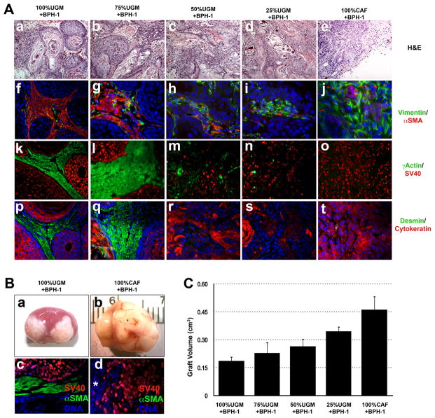
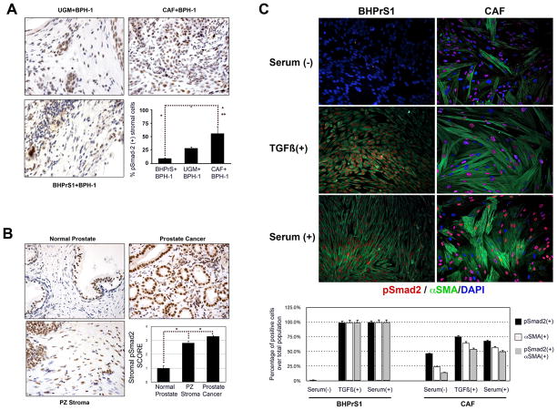
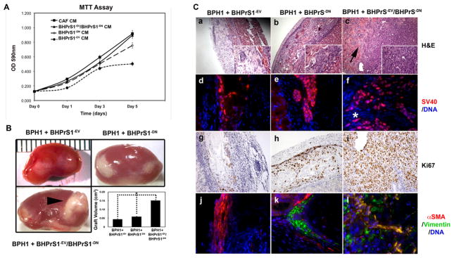
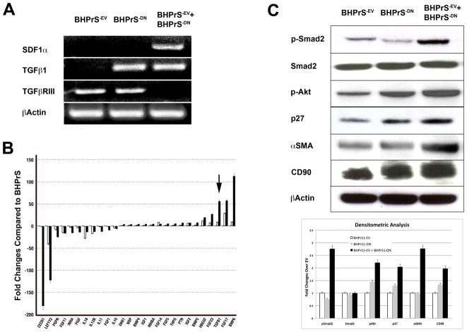
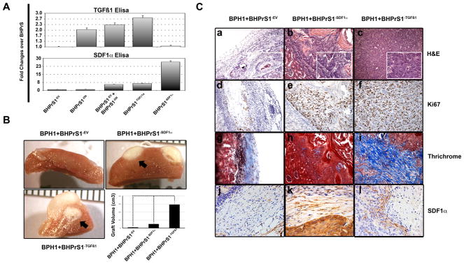
Carcinoma-associated fibroblasts (CAF) play a critical role in malignant progression. Loss of TGF-β receptor II (TGFβR2) in the prostate stroma is correlated with prostatic tumorigenesis. To determine the mechanisms by which stromal heterogeneity because of loss of TGFβR2 might contribute to cancer progression, we attenuated transforming growth factor beta (TGF-β) signaling in a subpopulation of immortalized human prostate fibroblasts in a model of tumor progression. In a tissue recombination model, loss of TGFβR2 function in 50% of the stromal cell population resulted in malignant transformation of the nontumorigenic human prostate epithelial cell line BPH1. Mixing fibroblasts expressing the empty vector and dominant negative TGFβR2 increased the expression of markers of myofibroblast differentiation [coexpression of vimentin and alpha smooth muscle actin (αSMA)] through elevation of TGF-β1 and activation of the Akt pathway. In combination, these two populations of stromal cells recapitulated the tumor inductive activity of CAFs. TGFβR2 activity in mixed stromal cell populations cultured in vitro caused secretion of factors that are known to promote tumor progression, including TGF-β1, SDF1/CXCL12, and members of the fibroblast growth factor (FGF) and bone morphogenetic protein (BMP) families. In vivo, tissue recombination of fibroblasts overexpressing TGF-β1 and SDF1/CXCL12 not only induced transformation of BPH1 cells, but also promoted a robust growth of highly invasive cells, similar to effects produced by CAFs. While the precise nature and/or origin of the particular stromal cell populations in vivo remain unknown, these findings strongly link heterogeneity in TGF-β signaling to tumor promotion by tumor stromal cells.
©2011 AACR.
Figures
Similar articles
-
Fibroblast growth factor-2 mediates transforming growth factor-beta action in prostate cancer reactive stroma.Oncogene. 2008 Jan 17;27(4):450-9. doi: 10.1038/sj.onc.1210663. Epub 2007 Jul 16. Oncogene. 2008. PMID: 17637743
-
Cross-talk between paracrine-acting cytokine and chemokine pathways promotes malignancy in benign human prostatic epithelium.Cancer Res. 2007 May 1;67(9):4244-53. doi: 10.1158/0008-5472.CAN-06-3946. Cancer Res. 2007. PMID: 17483336
-
Lack of transforming growth factor-β signaling promotes collective cancer cell invasion through tumor-stromal crosstalk.Breast Cancer Res. 2012 Jul 2;14(4):R98. doi: 10.1186/bcr3217. Breast Cancer Res. 2012. PMID: 22748014 Free PMC article.
-
Role of the stromal microenvironment in carcinogenesis of the prostate.Int J Cancer. 2003 Oct 20;107(1):1-10. doi: 10.1002/ijc.11335. Int J Cancer. 2003. PMID: 12925950 Review.
-
Role of stroma in carcinogenesis of the prostate.Differentiation. 2002 Dec;70(9-10):473-85. doi: 10.1046/j.1432-0436.2002.700902.x. Differentiation. 2002. PMID: 12492490 Review.
Cited by
-
Genes upregulated in prostate cancer reactive stroma promote prostate cancer progression in vivo.Clin Cancer Res. 2014 Jan 1;20(1):100-9. doi: 10.1158/1078-0432.CCR-13-1184. Epub 2013 Oct 22. Clin Cancer Res. 2014. PMID: 24150235 Free PMC article.
-
Fibroblast heterogeneity in prostate carcinogenesis.Cancer Lett. 2022 Jan 28;525:76-83. doi: 10.1016/j.canlet.2021.10.028. Epub 2021 Oct 29. Cancer Lett. 2022. PMID: 34715252 Free PMC article. Review.
-
Antidepressant fluoxetine and its potential against colon tumors.World J Gastrointest Oncol. 2014 Jan 15;6(1):11-21. doi: 10.4251/wjgo.v6.i1.11. World J Gastrointest Oncol. 2014. PMID: 24578784 Free PMC article. Review.
-
Enduring epigenetic landmarks define the cancer microenvironment.Genome Res. 2018 May;28(5):625-638. doi: 10.1101/gr.229070.117. Epub 2018 Apr 12. Genome Res. 2018. PMID: 29650553 Free PMC article.
-
Stromal matrix metalloproteinase 2 regulates collagen expression and promotes the outgrowth of experimental metastases.J Pathol. 2015 Apr;235(5):773-83. doi: 10.1002/path.4493. Epub 2015 Jan 5. J Pathol. 2015. PMID: 25469981 Free PMC article.
References
-
- Schor SL, Schor AM. Hypothesis: Persistent expression of fetal phenotypic characteristics by fibroblasts is associated with an increased susceptibility to neoplastic disease. Expl Cell Biol. 1987;55:11–7. - PubMed
-
- Schor SL, Schor AM, Rushton G. Fibroblasts from cancer patients display a mixture of both foetal and adult-like phenotypic characteristics. J Cell Sci. 1988;90:401–7. - PubMed
-
- Kalluri R, Zeisberg M. Fibroblasts in cancer. Nature reviews. 2006;6:392–401. - PubMed
-
- De Wever O, Demetter P, Mareel M, Bracke M. Stromal myofibroblasts are drivers of invasive cancer growth. Int J Cancer. 2008;123:2229–38. - PubMed
Publication types
MeSH terms
Substances
Grants and funding
LinkOut - more resources
Full Text Sources
Other Literature Sources
Medical
